Herbs

Lavender

Where it’s Found

Most common varieties of lavender are native of Mediterranean, Atlantic islands, Asia Minor and India, however, lavender can be grown nearly anywhere in the world. There are at least twenty-five species of lavender.

How to Process Lavender

Processing lavender begins with drying the flowers.

For small gardens, the flowers are harvested and bundled, hung and dried in a well-ventilated shady place. For medium-sized gardens, the flowers are harvested and placed flat on drying trays up off the ground. The best drying conditions places the tray in a well ventilated, shaded area. Larger fields are commercially processed with dryers.

The volatile oil is distilled from the dried flowers through a steam process.

Propagation of Lavender

Hardy Lavender’s best method of propagation is by cuttings or layering. Long cuttings from plant three years old or more have higher success. Select healthy branches of the early spring softwood, dip into root promoting powder and plant to a 3 – 4 inch depth.

To propagate over winter, many gardeners choose to layer several branches. Remove the leaves from the midsection of low-lying hardwood stems. Cover the midsection with dirt, leaving the ends above the soil.

Tender Lavenders propagate easy by seed. Start seeds indoors in a flat tray with good drainage. Fill the tray with one inch of soil, sift the seeds, cover seeds with ½ inch of sand. Water the soil with a fine spray. Keep soil moist. Cover with clear plastic wrap for 14 days until seedlings sprout. Once the seedlings have their first four leaves, the lavender is ready to transplant.

Cultivation

Notable similarities of all varieties is the shape and size of the flowers which range in color as they ripen from pale mauve, blue, blue-green, to vibrant purple flowers. The leaves are narrow width and grayish green color. The more loam in the soil increases foliage growth but does not enhance flower growth.

Lavender (Lavandula officinalis) is a perennial shrub with an average height of 32 inches at full bloom with an average circumference of 24 inches.

The plant grows best in well drained, poor, calcareous soil with full sun. With proper growing conditions, it’s possible to have two harvests; first in late spring, and second in autumn after a cool summer.

English Lavender (Lavandula vera)

This variety of lavender grows best in well drain soil. Since it grows in cloudy areas it usually isn’t ready for harvest until Autumn.

History

From the Latin “lavare” which means to wash or bathe, ancient laundresses added lavender to the wash water.

The earliest medicinal record for Lavender is among the first century writings of Dioscorides, a Greek physician who lived in Ancient Rome. His materia medica was in continuous use from his lifetime until 1600AD. This ancient doctor suggested lavender to treat “griefs of the thorax”. European folk tradition regarded lavender for use on wounds and as a remedy for worms. Victorian medicinal era used lavender to relieve regular headaches, migraines, trembling, passions and cramps.
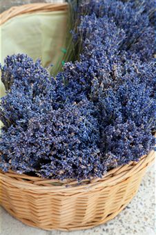
Lavender Benefits
Alchemists categorize lavender as hot gender under the ruling planet of Mercury. Used in concoctions that required representation of Air, lavender served as an ingredient for purification baths of Midsummer Celebrations as recounted in Shakespeare’s famous play.

Renaissance tales call lavender by the folk names “Spike” and “Elf Leaf”. During this period lavender gained a reputation for enhancement of love, specifically to attract men into love affairs.

Craft and home products use lavender as a main or important ingredient of the mixtures. Some of the products include: sachets, drawer liners, hot pads, wands, potpourri, tussie mussies, and wreaths.

Use

Lavender is regularly used as an anti-inflammatory, carminative, diuretic, insomnia, mild sedative, relaxant, stomachic, and spasmodic.

Since the earliest use of the steam distillation process developed in Persia during the 11th century lavender has been often used in fragrance recipes. In fact, lavender oil is a basic fragrance ingredient many popular personal care products and perfumes.

Traditional use of lavender in whole or in part helps increase healing of a variety of ailments. A sampling of the uses of lavender as treatments for ailments include: a topical application for athlete’s foot, an aromatherapy remedy for breastfeeding problems, a topical remedy for burns, an infusion for colds, a chest run for coughs, an aromatic for depression, in massage oil for headaches, a topical spray for an insect repellant, a topical ointment for treating stings, and a topical salve for sunburns.

Contemporary uses add to the traditional list of medicinal uses to aid healing of acne, cellulite, emotional heath, fibromyalgia, menopause, and stress.

Variations

In general, all lavender are perennial plants in garden growing zones 4 through 8. Older plants look good through winter with a silvery hue. Younger and new growth is brighter green than gray. Leaves vary in size and dimensions. Stems shoot up during early spring. The flowers also called spikes vary from a tapered shape to blunt ends.

Hardy Lavender

Hardy Lavenders are of a Mediterranean origin.

Dwarf Musthead named for the gardener who cultivated it. Dwarf Musthead is a low compact version of lavender with stems that spike at about 4 inches above the shrub. The spike flower is a true lavender purple. It can grow equally well in garden beds or in containers.

Folgate Lavender grows slightly larger than the Dwarf Musthead. Spike flowers are blue and grow 4 – 5 inches above the bush.

Grey Hedge, another variety of Lavandula angustifolia shrub or hedge grows larger with a silver hew to the leaf. The spikes mauve colored and are more pointed and thin.

Hidcote Purple is among the largest of the Lavenders growing to 30 inches with long dark purple spikes which gives the Hidcote Purple Lavender its name.

Old English Lavender is among the original English garden varieties of Lavandula angustifolia are hardy with their denser shrub with broader grey green leaves. The spike flower blooms a mauve color.

Seal Lavender grows to a height of a full 3 feet with the optimum growing conditions. The leaves are gray-green year-round. The flowers compliment the bush color with a blue-mauve flower. This variety of Lavandula angustifolia yields an extended bloom season up to four months.

Twickel Purple compares with the Hidcote Purple but in a smaller form. Twickel Purple is an unusual variety in that the spikes present in a fan-like formation of the deep mauve flowers.

Broad-leafed Lavender (Lavandula latifolia) has much broader gray colored leaves. Lavandula latifolia is the common commercial variety because it has a comparatively larger quantity of fragrant oil.

Dutch Lavender (Lavandula x intermedia) is the result of cross-breeding of Lavandula angustifolia and Lavandula latifolia. A distinguishing quality from the parent plants is the leaves are narrow compared to Lavandula latifolia but broader than Lavandula angustifolia. The spike flowers are long & branched. Dutch Lavender blooms later in summer and has a notably strong scent.

Wooly Lavender ( Lavandula lanata) grows to 2 feet with a diameter of up to 3 feet. This short squat version of lavender has gray leaves with tiny hairs on the stems and leaves. The spike flowers can grow to a foot above the bush with heads of dark blue.

Tender Lavenders

Tender Lavenders origins are traced to the Spanish & Southern French regions.

Lavandula stoechas is the historic lavender used by Romans during ancient times through to the Middle Ages. The green leaves have a pungent and notably camphorous scent. The spike flowers of this lavender are irregular globes with small flowers hidden between the purple bracts. The two long brilliantly purple bracts grow to a point reaching upward to 1 ½ inches in length. This variety grows well in acidic soils.

French Lavender (Lavandula dentata) has graceful green leaves. The fragrance of these locally known Spanish Lavender or French Lavender blends balsam with camphor. This variety blooms prolifically throughout most of the year.

Active Ingredients

The constituents of Common Lavender (Lavandula officinalis) consist in the form of volatile oil, tannins and coumarins. Within the Volatile oil up to 1.5% per volume, the active ingredients listed in degrees from highest to lowest in content are linabol, linalyl acetate, lavendulyl acetate, terpinenol, cineole, camphor, borneol, pinene, and limonene; within the coumarins, the active ingredients include coumarin, umbelliferone and hemiarin .

Spike Lavender (Lavandula latifolia) has higher percentages of camphor and cineole than other forms of lavender.

Who uses

Popular in French Cuisine, a chef may use lavender in a recipe or as a part of a sprinkle on top of savory dishes including beef or fish, or on cookies or pastries.

Natural Cosmetologist who seek to enhance not only their clients beauty but also their health work with natural botanicals that have low allergens and high health value. Lavender as an edible as well as its topical usage is perfect for the whole body aware cosmetologist.

Massage Therapist enhance the massage experience by providing just an bit more relaxation by using essential oils in their massage oil. Lavender scent helps relax the mind, as the lavender oil works its way in through the skin to relax the muscles as well. Aroma therapists use lavender as a basic scent in many of their mixtures. The relaxing aroma can improve relaxation, and increase restful sleep as well as boost pleasant and rejuvenating dreams.
Lavender Uses
Traditional Folk Doctors used lavender for a variety of ailments. It seems that lavender was among the stock medicines before allopathic medicine developed.

From flesh wounds or burns to remedies for breathing problems or headaches, lavender was a value herb since the beginning of medicinal treatments.

Herbalists & midwives used lavender to aid healing before, during and after the birth process. Lavender’s qualities helped reduce stretch marks, aided in stretch the perineum and removed the smells of birth by adding lavender to the bedding.

Crafters use lavender in a variety of products for the house. From front door wreaths to drawer sachets the Victorian housewife or 21st Century crafters adore lavender for the variety of colors and the powerful and lingering scent.

Popular Brands/Forms

Aroma therapy oil created from a mixture of essential oil of lavender with base oil. Popular brands: Ananda Apothecary, Aura Cacia.

Flowers are used whole, crushed or ground in culinary or medicinal mixtures. Popular brand for culinary bulk flowers: Starwest Botanicals, Frontier Herb.

Cosmetics used lavender in beauty products including: facial scrub, masks and other astringent mixes. Lavender steam for complexion treatment leaves skin moist and firm. The steam is also good for the lungs. Popular brands: Little Mama Products, Avalon Organics.

A popular scent, lavender can be found in handmade soaps, shampoos, conditioners, deodorants and other personal care products. Popular brands: South of France, Kiss My Face.

Typical Dosage & Usage Regime

Traditional use and dose of lavender flowers for cough and colds suggests brewing an infusion of the flowers and drinking several cups of lavender throughout the day. Another way to administer lavender for cold symptoms is through steam inhalation two or three times per day to loosen phlegm and relax bronchial passages.

Medicinal Herbalists of the 19th century used of the higher potency lavender oil in the treatment of diphtheria, streptococcus and typhoid bacteria. Massage therapists add one or two drops of lavender oil to blank massage oil to help increase muscle relaxation, aid in treatment of headaches, neuralgic and rheumatic pain.

Medicinal infusions are made as a tea. Place the herb in a serving teapot. Pour boiling water from a kettle over the herb into the teapot. Place the lid on the teapot to steep the herb for 10 minutes. Strain the herb as the infusion is poured into a cup. Ratio: 1-2 teaspoons per cup of water.

  • Medicinal Extract Ratio: 10-30 drops in water 1-4 times per day, as directed by a practitioner.
  • Medicinal Tincture: 5 ml, twice daily for depression or headaches.
  • Chest Rub Ratio: Add 1 ml oil and 5 drops chamomile oil to 10 ml base oil for bronchial spasm.
  • Massage Oil Ratio: 1 ml lavender oil to 25 ml base oil.

Whole or ground flowers are used in a variety of culinary dishes including: lavender cookies, lavender sugar, lavender lemonade, lavender vinegar, lavender honey, lavender jelly.

Potential Side Effects

FDA has not tested evaluated or approved any form of Lavender for use. Typical Cautions for Lavender as with all other herbs known as relaxants include: caution while operating vehicles, talk to your doctor before use if you may be pregnant or have allergies or drink alcohol or are taking any mediations especially antihistamines and sedatives.

Cautions include warning against overdose but there is no known quantity identified as excessive. Lavender as a relaxant leads suspicions to include drowsiness as a symptom of an overdose.

More likely, identified side effects can be attributed to plant-based allergies. If you have a history of plant allergies approach lavender with normal caution. Allergic reactions include respiratory or dermatologic symptoms including difficulty breathing, swelling of the throat or facial areas or hives, including itching, redness and swelling. Should these symptoms manifest follow regular allergy protocol.

Typical Costs

Current average price of dried flowers is $10-$20 per pound.

Current average price of lavender oil is $9.00 – $12.00 for a one ounce bottle; $80.00 – $95.00 for a 16 ounce bottle.

Where Found Online & Offline

Local health food stores, culinary markets and herbs stores generally stock lavender or can order it for you.

Use reputable suppliers when buying online. It’s better to purchase lavender from suppliers with excellent reputations such as Dry Creek Herb Farm in Sacramento, CA http://drycreekherbfarm.com/catalog/ or Jean’s Greens in Upstate New York http://www.jeansgreens.com/.

Antioxidants

Tocopherols

Tocopherols (Vitamin E)

Vitamin E is a vitamin that is fat-soluble and comes with antioxidant properties. There are eight different forms in which vitamin E may exist:

  • Alpha tocopherol
  • Alpha tocotrienol
  • Beta tocopherol
  • Beta tocotrienol
  • Gamma tocopherol
  • Gamma tocotrienol
  • Delta tocopherol and
  • Delta tocotrienol

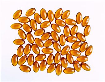
The most active form of vitamin E found in humans is the alpha-tocopherol, and it is for this reason that when daily allowance and dosing recommendations they are made, they are usually made in Alpha-Tocopherol Equivalents, or ATE. Vitamin E supplements may be found in synthetic or natural forms, the latter of which tends to be labeled with a “d,” such as in d-gamma-tocopherol. The synthetic forms, on the other hand, tend to be labeled with “dl,” such as in dl-alpha-tocopherol.
Tocopherol Information

Treating Different Health Conditions

Vitamin E has often been proposed as part of a treatment or prevention plan for a number of different health conditions, usually due to its antioxidant properties.

However, outside of treating the rare vitamin E deficiency, there is no scientifically proven medicinal usage of the supplementation of vitamin E outside of maintaining the recommended dosage. Ongoing research is current, especially in treating heart disease and cancer.

There have been recent concerns associated with how safe it is to take high doses of vitamin E supplements. The idea has been proposed about whether there is an increased risk of bleeding, especially with those who are also taking aspiring, heparin, warfarin and other blood thinners, or in patients who are deficient in vitamin K.

Evidence that has recently come to light has come to light that taking high doses of vitamin E supplements might increase the risk of death from “all causes,” though a separate study said there was no increase towards mortality for women who supplemented vitamin E on a daily basis. Caution is warranted.

Uses Based on Tradition or Theory

The following list are uses that are based on scientific theories or traditions. More often than not, they are not fully tested in humans, meaning that the effectiveness and safety are not necessarily proven. Some conditions are potentially very serious and should only be evaluated by a qualified professional.

  • Abortifacient
  • Acne
  • Aging prevention
  • Aging skin
  • Air pollution protection
  • Allergies
  • Amiodarone pulmonary toxicity prevention
  • Bee stings
  • Benign prostatic hypertrophy
  • Beta-thalassemia
  • Blood disorders (porphyria)
  • Breast pain or inflammation (mastitis)
  • Bronchopulmonary dysplasia in infants born prematurely
  • Bursitis
  • Cardiomyopathy
  • Celiac disease
  • Chemotherapy extravasation
  • Chorea, or chronic progressive hereditary
  • Congestive heart failure
  • Crohn’s disease
  • Cystic fibrosis
  • Dermatitis
  • Diaper rash
  • Digestive enzyme deficiency
  • Doxorubicin hair loss
  • Duchenne muscular dystrophy
  • Dyspraxia
  • Energy enhancement
  • Exercise recovery
  • Frostbite
  • Gastric ulcer
  • Granuloma annulare
  • Hair loss
  • Heart attack
  • Heart transplant rejection
  • Hereditary spherocytosis
  • Huntington’s disease
  • Hypertension
  • Impaired glucose tolerance
  • Impotence
  • Leg cramps
  • iver disease
  • Liver spots
  • Lung cancer
  • Male fertility
  • Menopausal symptoms
  • Menstrual disorders
  • Miscarriage
  • Mucositis
  • Muscle strength
  • Myotonic dystrophy
  • Neuromuscular disorders
  • Nitrate tolerance
  • Oral leukoplakia
  • Labor pain
  • Pancreatitis
  • Peptic ulcers
  • Physical endurance
  • Poor posture
  • Post-angioplasty restenosis
  • Post-operative recovery
  • Pre-eclampsia
  • Radiation induced fibrosis
  • Reperfusion injury protection
  • Restless leg syndrome
  • Sickle cell
  • Skeletal muscle damage
  • Skin damage
  • Skin disorders
  • Sperm motility
  • Sunburn
  • Thrombophlebitis and
  • Ulcerative coloitis
  • Dosing

The doses as provided in this section are based on research by scientists, traditional use, publications or expert opinion. There are many supplements and herbs that have not been fully tested, meaning the effectiveness and the safety of taking them are not necessarily proven. Some brands may differ in how they make their supplements, using other ingredients, even with its own brand. They may not apply to all sorts of products. Always read the product labels.

Dietary Sources

The following foods are known to contain vitamin E:

  • Eggs
  • Fortified cereals
  • Fruit
  • Green leafy vegetables
  • Meat
  • Nuts
  • Nut oils
  • Poultry
  • Vegetable oils
  • Argan oil
  • Olive oil
  • Wheat germ oil and
  • Whole grains

Cooking may destroy some of the vitamins.

Adults

It is believed that most adults within the United States already have a sufficient amount of vitamin E from their dietary sources. The recommended allowance for adults over the age of 14 years is 15 milligrams ATE. The recommended allowance for pregnant women of any age whatsoever is about 15 milligrams ATE of vitamin E per day, with breastfeeding women of any age whatsoever needing 19 milligrams per day.

Children

Children between the ages of one and three years is six milligrams ATE per day. For children between the ages of four and eight years is seven milligrams ATE per day. For children between the ages of nine and 13 years is 11 milligrams ATE per day. Anybody over the age of 14 years is to receive 15 milligrams ATE of vitamin E per day. Pregnant women of any age at all should receive 15 milligrams ATE per day, and those who are breastfeeding, regardless of the age, should obtain about 19 milligrams ATE of vitamin E per day. No amount has been approved for infants.

Safety

It is not the position of the United States Food and Drug Administration, or FDA, to approve the regulation of supplements and herbs. Therefore, the effects may vary, there is no guarantee for the purity or the safety of the products, and there is no guarantee about its strength.
Vitamin E
For this reason, it is vital to always read the labels on the product. For those who suffer from a medical condition or are taking any other supplements, herbs or drugs, it is important to go see and consult with a qualified professional before taking on a new therapy. Also make sure to consult a doctor at once if experiencing any side effects.

Side Effects and Warnings

Evidence that has recently come to light has come to light that taking high doses of vitamin E supplements might increase the risk of death from “all causes,” though a separate study said there was no increase towards mortality for women who supplemented vitamin E on a daily basis. However, these results are often criticized by experts who said that they are based on recalculations of results from earlier studies of mixed quality and with variable results. However, it is still currently the best evidence available and as it stands everybody should take caution with taking vitamin E.

It is considered to be safe to take vitamin E supplements for a short period of time, only up to the recommended upper intake level. However, vitamin E has the potential of being unsafe when doses are taken orally at levels exceeding that of which is recommended. The RDA obtained through food consumption is generally thought to be both safe and beneficial.

Skin Reactions

Contact dermatitis and other kinds of skin reactions have previously been reported after using topical vitamin E preparations, including deodorants that have vitamin E in them or ointments. Rarely, it has been shown that vitamin E supplements can cause diarrhea, abdominal pain, diarrhea, nausea, or symptoms similar to that of influenza. There also seems to be an increased risk of developing necrotizing enterocolitis when taking a large dose of vitamin E.

In rare cases, there has been associations between the supplementation of vitamin E and gonadal dysfunction, as well as diminished kidney function.

Overdosing

Taking a high dosage of vitamin E on a regular basis is also believed to increase the risk of bleeding, and this is because the vitamin inhibits platelet aggregation and there is the antagonism of clotting factors dependent on vitamin K, especially with those who are deficient in the vitamin. Also associated with vitamin E supplementation, though rare, are fatigue, headache, dizziness, blurred vision and weakness.

People who suffer from retinitis pigmentosa should not consider taking vitamin E orally because it does not seem to slow down the decline of vision, and it actually seems to be associated with losing visual activity on a more rapid basis. This theory, however, has been questioned.

Pregnancy and Breastfeeding

There are numerous prenatal vitamins that are known to have small levels of vitamin E in them. It may be preferred to take natural kinds of vitamin E rather than the forms that come synthetically.

It is not recommended to take more vitamin E than the level of Recommended Dietary Allowance states that otherwise healthy pregnant women should have. Outside of this recommendation, there is currently no sufficient evidence that shows the safety levels of taking a higher dosage of vitamin E, whether injected, topical or oral, throughout the duration of breastfeeding and pregnancy. Therefore, it is not recommended to exceed such recommended dosages.

Vitamins

B12

B12

(Cyanocobalamin, Hydroxocobalamin, Methylcobalamin, Cobamamide)

Vitamin B12 is an essential vitamin that is water soluble. It is also very commonly found in numerous different kinds of foods, such as dairy products, fish, meat and shellfish. Frequently, vitamin B12 is used in conjunction with other vitamin B types in a complex formulation.

The vitamin serves to keep nerve cells healthy, as well as the red blood cells, and it is also needed in order to make DNA, which is the genetic material that is found in every cell. Vitamin B12 is bound to food by way of the protein. During digestion, the stomach releases the B12 from protein using its own hydrochloric acid, and upon its release, it mixes into the intrinsic factor, another substance, prior to being absorbed into the blood.

B12 Deficiency

It is extremely rare to suffer from vitamin B12 deficiency because the human body works to store several years worth of the nutrient. As such, those who are elderly are the ones who are the greatest risk to experiencing deficiency. However, deficiency is also possible when a person is not able to use the vitamin.

Pernicious anemia is an example of a type of disease that makes it impossible for a person to absorb this essential vitamin from their intestinal tract. Furthermore, a vegan or a strict vegetarian may be prone to a state of deficiency if they are not taking in the correct dosages of B12.

A person can easily get a full day’s worth of vitamin B12 simply by eating a cup of milk with a cup of raisin bran, or a chicken breast with a hardboiled egg and a cup of yogurt, plain and low in fat.

Synonyms

Vitamin B-12 is also known as:

  • B-12
  • B Complex
  • B Complex Vitamin
  • Bedumil
  • Cobalamin
  • Cobalamins
  • Cobamin
  • Cyanocobalamin
  • Cyanocobalaminum
  • Cycobemin
  • Hydroxocobalamin
  • Hydroxocobalaminum
  • Hydroxocobemine
  • Idrossocobalamina
  • Methylcobalamin
  • Vitadurin and
  • Vitamin B-12.

Evidence of Successful Vitamin B12 Therapy

The following uses have been previously tested in animals or humans however, neither the safety nor the effectiveness have always been proven in these studies.

Vitamin B12 Deficiency

Previous scientific research have shown that when a person has a deficiency of the vitamin B12, it may lead to complications such as abnormal psychiatric and neurologic symptoms. Some of these such symptoms in question may include:

  • Muscle weakness
  • Ataxia, which is shaky movement and an unsteady gait
  • Hypotension, which is low blood pressure
  • Incontinence
  • Spasticity
  • Mood disturbances
  • Vision problems
  • Psychoses and
  • Dementia.

Researchers say that the previously mentioned symptoms have a tendency to occur even when the levels of the vitamin B12 are only a little bit lower than they are supposed to be, and when they are considerably higher than levels that are considered to be normal for those with anemia.

People who are at risk for developing deficiency of vitamin B12 include those who:

  • are strict vegetarians
  • are elderly
  • are breastfed infants

are people who need more vitamin B12 due to pregnancy, hemolytic anemia, thyrotoxicosis, hemorrhage, liver disease, malignancy or kidney disease.

Uses Based on Tradition or Theory

The uses as described below are based upon scientific theories and tradition. Caution is warranted because not every supplement or herb has been fully and extensively tested, meaning that the effectiveness and the safety thereof has not yet been proven. It is important to read the labels on the products, as well as to discuss the dosages with a qualified provider of healthcare prior to starting any new therapy.

  • Aging
  • AIDS
  • Allergies
  • Amyotrophic lateral sclerosis
  • Asthma
  • Autism
  • Chronic fatigue syndrome
  • Cognitive function
  • Depression
  • Depressive disorder
  • Diabetes
  • Diabetic peripheral neuropathy
  • Energy level enhancement
  • Growth disorders
  • Hemorrhage
  • Immunosuppression
  • Improving concentration
  • Inflammatory bowel disease
  • Kidney disease
  • Liver disease
  • Male infertility
  • Malignant tumors
  • Memory loss
  • Mood
  • Mouth and throat inflammation, or atrophic glossitis
  • Multiple sclerosis
  • Myoclonic disorders, or spinal myoclonus
  • Neural tube defects
  • Osteoporosis
  • Peridontal disease
  • Poisoning by cyanide
  • Protection from smoke by tobacco
  • Psychiatric disorders
  • Seborrheic dermatitis
  • Seizure disorders by way of West syndrome
  • Tendonitis
  • Thrombosis
  • Thyrotoxicosis
  • Thyroid storm, or adjunct iodides
  • Tinnitus
  • Tremor
  • Vitiligo
  • Dosing

The below figures are solely based on traditional use, scientific research, expert opinion and publications. Caution is warranted because not every supplement or herb has been fully and extensively tested, meaning that the effectiveness and the safety thereof has not yet been proven. Each brand will probably be made differently, even within its own brand, using a variety of different ingredients. Not all brands may make their products to match the below dosages, so it is important to read the labels on the products, as well as to discuss the dosages with a qualified provider of healthcare prior to starting any new therapy.

Adults (over 18 years old)

The recommended dietary allowance for any adult and adolescent over the age of 13 is 2.4 micrograms on a daily basis. Pregnant females, whether adult or adolescent, should take 2.6 micrograms on a daily basis. Females who are lactating, whether adult or adolescent, should take 2.8 micrograms on a daily basis.

Because, more often than not, 10 – 30 percent of elderly people do not efficiently absorb enough vitamin B12 through food consumption, it is recommended for those over the age of 50 years to meet their recommended dietary allowance by way of eating foods that are fortified with B12 or by way of taking a vitamin B12 supplement.

To maintain the levels of vitamin B12 in elderly people, they should consider taking 25 – 100 micrograms of supplementation per day. Other indications will require the consultation of either a doctor or a pharmacist. Vitamin B12 may be taken by way of the mouth or provided by a qualified professional in health care and by way of intramuscular injection.

Children (under 18 years old)

Not all pediatric age groups have had recommended dietary allowances established for them. For this reason, there are instead Adequate Intake levels used. Infants are between the ages of newborn and six months should only have 0.4 micrograms. Infants between the ages of seven months and twelve months should take 0.5 micrograms.

Children between the ages of one and three years should take 0.9 micrograms. Children between the ages of four and eight years should take 1.2 micrograms. Children between the ages of nine and 13 should take 1.8 micrograms.

Safety

The United States Food and Drug Administration does not typically regulate supplements and herbs. As such, there are no guarantees with regards to the purity, strength or the safety of the products, and the effects will likely vary. Therefore, it is important to read the labels on the products. Consult a doctor if there is a medical condition present or if any other supplements, herbs or drugs are being taken, especially when considering new therapy. Consult a doctor if any side effects occur, especially as described below.

Allergies

Anybody who is sensitive or allergic to colbat, cobalamin or any other kind of product ingredient should avoid taking any vitamin B12 supplements.

Side Effects and Warnings

Patients who are undergoing angioplasty are advised to take caution with vitamin B12. This is because an intravenous loading dose of vitamin B12, vitamin B6 and folic acid on a daily basis after coronary stenting has shown to potentially increase the rate of restenosis. Because of the risk of harm, it is recommended not to take this combination of vitamins for those who are obtaining a coronary stent.

Other side effects have been reported from taking vitamin B12, such as transitory exanthema, itching, rash, diarrhea and urticaria. Cases of rosacea fulminans have been associated with vitamin B12 and pyridoxine, which is hallmarked by erythema with nodules, pustules and papules. These symptoms have the potential to persist upwards of four months once the supplement usage has been stopped, and it may need to be treated using topical therapy and systemic corticosteroids.

Peripheral vascular thrombosis is another side effect to be reported. By treating a deficiency of vitamin B12, it is possible to unmask polycythemia vera, hallmarked by an increase of how many red blood cells there are, and it increases the volume of blood. Attempting to correct megaloblastic anemia using vitamin B12 in susceptible individuals may lead to the onset of fatal hypoklameia or gout. Additionally, it might obscure the folate deficiency of megaloblastic anemia, so caution is warranted.

Pregnancy and Breastfeeding

It is likely safe to take vitamin B12 orally provided pregnant and breastfeeding women do not exceed the dietary allowance that is recommended for them to take. Currently, there is an insufficient amount of information available with regards to the safety of taking a larger amount of vitamin B12, so it is not recommended.

Conditions

Endometriosis

Endometriosis is an illness that affects women of childbearing age with debilitating pain and infertility. Endometriosis causes multiple symptoms that can be difficult to manage, diagnose, and treat. It is thought to affect 5-10% of pre-menopausal women in the United States.

Alternative Names for Endometriosis

The name endometriosis comes from the Latin form of the words “endo,” meaning “inner;” the portion of the word “metri” refers to the uterus or womb; and “-osis” means “condition.” Although the name describes the adverse condition of the uterus, endometriosis can also affect surrounding structures within the pelvis and abdominal cavity.

Endometriosis has also been called by other names that refer to the unusual results of the disease itself. It has been named adenomyosis, which is a type of endometriosis in which uterine lining implants and grows within the muscular walls of the uterus itself. Endometriosis may also be referred to as chocolate cysts or endometriomas, both of which are by-products of the disease.

Chocolate cysts are pockets of blood that are found growing outside the uterus within the pelvic cavity. Because the blood inside the cyst is old, it has a dark appearance, similar to that of chocolate. Endometriomas are another name for growth of endometrial tissue found in various locations throughout the abdominal and pelvic cavity.

Symptoms of the Disease

Symptoms of endometriosis are similar but may vary in severity. Some women may suffer from extreme pain and heavy bleeding with their menstrual periods. Other women may be unaware that they are affected by endometriosis until they receive a diagnosis of infertility. The following symptoms are indications of endometriosis:

  • Pelvic pain
  • Heavy bleeding with menstrual periods
  • Spotting or bleeding between periods
  • Pain with urination
  • Pain with bowel movements
  • Infertility
  • Gastrointestinal symptoms, such as diarrhea or constipation
  • Pain with intercourse
  • Fatigue

Pelvic pain is the most common sign for women experiencing endometriosis. Pain may be located within the pelvic area, near the uterus, or it may radiate to the back. Some women feel pain near the location of their ovaries; it may radiate into the abdomen, down the legs, or up into the shoulder. The pain can be a constant dull ache, sharp and intermittent, or a combination of both. The severity of pain varies with each woman, some experiencing acute pain that is debilitating. Others suffer from a constant, dull ache. A percentage of women experience no pain with endometriosis.

Endometriosis can cause excessive bleeding during a woman’s menstrual period, resulting in heavy flow. The period may last significantly longer than average, continuing many days or even weeks at a time. Menstrual flow may have large clots and be associated with severe uterine cramping. A heavy menstrual flow is difficult for many women to manage and can be extremely uncomfortable.

Timetable & Menstrual Cycle

The end of a menstrual period typically indicates that approximately a month will pass before starting another. For the patient with endometriosis, there may be spotting of blood or recurring bleeding similar to menstrual flow but lasting for a shorter duration. This occurs mid-cycle and may be associated with menstrual symptoms, such as cramps. This can be challenging to manage for many women, as they may be unprepared for breakthrough bleeding mid-cycle.

Some women with endometriosis experience pain with urination. Endometrial tissue may migrate from the inside of the uterus to the bladder or ureters and implant itself. The action of emptying the bladder can then cause pain during urination or during the sensation of having to void. There may be small amounts of blood that is seen in the urine.

Bowel Movement Issues

Pain during a bowel movement is an associated symptom of endometriosis, as the bowel and rectum become locations for the implantation of endometrial tissue. The pain and bleeding at these sites results in pain during movement of the bowels. A woman may also experience small amounts of blood in the stool.

Infertility & Female Issues

Infertility is closely associated with endometriosis. For women that do not experience pain or bleeding symptoms that are typical of the disease, infertility is often the source of diagnosis. There can be multiple sources of infertility for the woman suffering from endometriosis. The implantation of endometrial lining in various parts of the pelvic cavity leads to bleeding and scar tissue formation that can block the process of fertilization. For example, scar tissue build-up within the fallopian tubes can prevent a released egg from finding its way to the uterus for fertilization.
Endometriosis Herbal Remedies
A hormonal imbalance is often associated with endometriosis, and in order to achieve pregnancy and successfully grow a baby to term, a delicate balance of hormones must remain in the body. Endometriosis involves an imbalance that may prevent implantation of a fertilized egg into the uterus, or the growth of the egg into a fetus, resulting in miscarriage.

Some women experience gastrointestinal symptoms such as diarrhea or constipation. Lesions of endometrial lining may localize on the bowel or intestine, causing a change in bowel habits and producing loose stools or reducing the frequency of bowel movements.

Endometrial lesions may occasionally appear in or near the vagina or cervix of a woman, or they may deposit in the peritoneal cul-de-sac. The cul-de-sac is the area located between the back wall of the uterus and the rectum. Endometrial lesions found in these locations can cause pain during sexual intercourse.

Fatigue

Fatigue is the result of several symptoms of endometriosis, including management of chronic pain and blood loss. Any chronic disease that leaves a patient handling negative symptoms such as pain can become exhausting. The body utilizes some of its own resources to respond to pain, but after an extended period of time, the body’s energy may become depleted, resulting in fatigue.

Large amounts of blood loss through menstruation or internal bleeding from lesions can also result in fatigue. For women with exceptionally heavy amounts of bleeding, laboratory tests such as an iron panel should be checked for possible anemia.

The management of chronic disease also leads to the possibility of depression for women with endometriosis. Living with constant pain, as well as experiencing discomfort during typically normal activities can be exhausting. Infertility for a couple with a desire to have a child can be a heartbreaking event, leaving them hoping for a baby but feeling depressed because of the illness. Women diagnosed with endometriosis should consider the possibility of depression as a potential long term effect of the disease and seek treatment if necessary.

Causes of Endometriosis

There is not one exact cause of endometriosis, but several theories have developed, all of which have research to back up some of their ideas. The process of menstruation typically develops as a monthly episode where the body releases an egg from one of two ovaries located near the uterus. The ovaries are connected to the uterus with the fallopian tubes, each a passageway for the egg to travel as it moves toward fertilization.

During the time preceding ovulation, the uterus has built up a healthy amount of interior lining made of blood and tissue that will provide a place for a fertilized egg to implant and then grow into a fetus. Without fertilization, the egg is shed along with this endometrial lining approximately once a month during menstruation.

Ovulation

The entire process of ovulation through menstruation is managed by specific hormones within the body. Two common hormones, called estrogen and progesterone are responsible for many of these factors, such as the maturation of an egg before ovulation and the development of endometrial lining in the uterus.

Endometriosis occurs when some of the endometrial lining that is normally found in the uterus migrates to other parts of the pelvic cavity and implants itself on other structures. These endometrial lesions can be found on many parts of the body, including the ovaries, the fallopian tubes, the bowel, the bladder, the ureters, or the wall of the peritoneum.

Lesions

In rare cases, lesions have been found on the diaphragm and even the lungs of some women. Although the lesions implant on areas outside the uterus, during the process of menstruation, the lesions bleed just as if they were still inside the uterus. It is this internal bleeding that causes pain and the multiple symptoms associated with endometriosis.

The reasons for the wandering endometrial tissue are unclear, but are thought to be related to one of the hormones affecting menstruation: estrogen. Increased amounts of estrogen have impacted the quantity of endometrial tissue found in other parts of the body, and is responsible for the monthly bleeding associated with the lesions.

Retrograde Menstruation

Another theory that may be a source of endometriosis is the concept of retrograde menstruation. In this situation, the body proceeds through the menstrual cycle as usual, but when it is time for the interior lining of the uterus to be shed, some of the blood and tissue travels up into the fallopian tubes and toward the ovaries where it exits into the pelvic cavity. This then allows some particles of the endometrial lining to attach to various internal organs and continue the process of endometriosis.

A genetic factor has also been attributed to the development of endometriosis. The risk of a woman developing the disease is ten times greater if she has close family member, such as a mother or sister, with endometriosis. The role of hormones in the process of heredity contributes to some of the changes that occur during the menstrual cycle, giving women that are direct relatives related menstrual symptoms, and therefore a genetic probability of disease development.

Risk Factors

Risk factors for developing endometriosis are related to some of the causes of the disease. Endometriosis is primarily a disease that affects women during their childbearing years, although it can also be found in post-menopausal women. Because of this, a woman of childbearing age (between menarche and menopause) is at a greater risk of developing endometriosis. Women that have a close female relative with the disease are also at greater risk. In addition, those women that take estrogen supplements are at risk due to the presence of the hormone related to the disease.

Prevention

There is little that can be done to prevent endometriosis, especially for women that are unaware that they have the illness. Endometriosis is a disease that is treated after symptoms appear. Awareness seems to be the highest form of prevention, as well as taking care of the body. Women should be aware of the risk factors associated with endometriosis and monitor their own body system for signs of the disease.

For women that suffer from heavy periods, vitamin supplements that contain iron may be effective in handling fatigue. The use of hormone supplements should be well monitored by a physician to determine if the body may be developing a hormone imbalance.

Diagnosis

Diagnosis of endometriosis is definitively made only by surgery. A physician can recommend surgery if a patient presents with the symptoms of endometriosis that is not managed by pain control or if she desires a pregnancy. A doctor should take a complete history of the woman, including menstruation, number of pregnancies, sexually transmitted diseases, and sexual history. The physician may ask about family members with endometriosis to find a possible genetic link. Lab work may be ordered to check for estrogen and progesterone levels.

If a woman is seeking treatment for infertility, a physician may attempt to treat the condition before performing surgery to rule out endometriosis. This typically occurs if infertility is one of the only symptoms. Infertility procedures, such as the administration of medications to induce ovulation, as well as intrauterine insemination may be performed in an attempt to achieve pregnancy first.

Hysterosalpingogram

A physician may perform a procedure called a hysterosalpingogram (HSG), which is a test to determine if the fallopian tubes are clear. If a woman is presenting with infertility and may have endometriosis, lesions may have appeared within the fallopian tubes, causing blockage or scar tissue that can prevent an egg from reaching the uterus for fertilization. During an HSG, a doctor administers a solution into the cervix of the patient where it travels up into the uterus, through the fallopian tubes, and into the ovaries.

The solution can be seen on x-ray and the physician is able to take radiographic images of the process to see the flow of fluid and determine if the fallopian tubes may be blocked. A blockage does not indicate definitively that endometriosis is present, but it can explain a reason for infertility, giving the doctor a reason to look for other possible signs of endometriosis.

Surgical Procedures

Surgical procedures to diagnose endometriosis include a laparoscopy to look for lesions in the pelvis. The doctor makes several small incisions in the abdomen and inserts a tube with a lighted camera on the end into the pelvic cavity. This enables the physician to look for lesions on organs and throughout the pelvis. If lesions are found, they can then be removed. The use of laparoscopy gives a physician a definite diagnosis of endometriosis and can help control some pain of the disease following the procedure.

Endometriosis is diagnosed according to stages, which depends on the severity of lesions, not the associated symptoms. Stage one is considered minimal, and there are very few lesions found outside the uterus. Stage two is referred to as mild endometriosis. There are more lesions and they are found deeper within the tissue.

Scar Tissue & Painful Side Effects

There is also the presence of scar tissue. Stage three is moderate endometriosis, which includes everything found in stages one and two, as well as implantations found on the ovaries. Stage four is inclusive of all stages, with large lesions and significant amounts of scar tissue. Stage four is considered severe endometriosis.

Pain and symptoms are not part of the diagnosis of which stage the disease is in. A woman may have severe pain, but with minimal lesions. Alternatively, a woman may present with only a diagnosis of infertility and no other symptoms yet have a diagnosis of stage four endometriosis with extensive scar tissue.

Treatment

Treatment of endometriosis is based on the stage of life of the woman and the severity of symptoms. For the childbearing woman who wishes for a future pregnancy, symptoms are treated through pain medication and other comfort measures. For example, pelvic pain can be helped with the use of anti-inflammatory medications and a heating pad to reduce discomfort. Surgery to remove lesions and scar tissue is also an option for the woman suffering severe symptoms but who still plans to have children. Surgery can reduce pain and bleeding associated with the disease and retains the internal structures needed for pregnancy.

Women that do not wish for a pregnancy have other options for treatment beyond comfort measures. The use of hormone therapy, particularly that found in oral contraceptives can reduce symptoms of pain and regulate menstrual bleeding. Progesterone is a hormone that can counterbalance excessive estrogen, the cause of many endometriosis symptoms.

Surgery Options

Surgery can also be an option for painful symptoms. Beyond laparoscopy to remove lesions and scar tissue, there is an option of removal of the uterus, fallopian tubes, and ovaries. This provides relief from excessive menstrual bleeding and can help with chronic pain, but is an invasive option that is reserved for the woman with particularly difficult symptoms.

Endometriosis can be a difficult disease to detect and to treat. Millions of women suffer from this disease throughout their childbearing years, making conception difficult and monthly periods unmanageable. Through the help of a physician, women suffering from endometriosis can be accurately diagnosed, and depending on their stage of life and desires, can be treated and find relief from these difficult symptoms.

Conditions

Chafing

Chafing is a very common problem that touches almost all people. Chances are that nearly everybody is familiar with what chafing is and has experienced it at some point in life. Though it is very annoying to have to deal with chafing, it is more of a discomfort than a serious medical problem in most cases. It is relatively simple to prevent chafing and even easy to remedy once it has become a problem. It is very rare that chafing seriously harms anyone.

By definition, chafing is almost more of a symptom of other problems than it is its own medical dilemma. It is a symptom of diaper rash, jock itch, and many other rashes. However, chafing is a problem in and of its own right for many people, and is not as severe as the rashes and conditions it is a symptom of. Chafing is the rubbing of the skin against something (clothing, other skin, etc.) in such a way as to cause friction. This action wears down on our outer-most protective layer of skin and it becomes sore and irritated as a result. Luckily, this is as easy to prevent as it sounds!

Symptoms

Symptoms of chafing may include skin that has turned pink or even red in more severe cases. Chafing usually becomes suddenly apparent to those affected via a burning sensation or stinging around the chafed area. The skin that has been subject to chafing will very likely be flakey and worn thin at the least and worn off entirely in the worst cases.

Causes

The cause of chafing is easy to understand. Basically, any kind of rubbing against the skin can irritate it and thus create chafing. This means there are many different things that can lead to chafing.

Literally any activity that calls for constant skin-on-skin rubbing can cause chafing. The same goes for any activity that causes the skin to rub against an object or repeatedly be rubbed by the fabric of clothing. The possibility for and severity of chafing only becomes worse when moisture is added.

Risk Factors

Those who are most obviously at risk for chafing are the highly physically active and the overweight, two groups who would normally have not much in common. In the case of athletes, any kind of poor fit in uniform can cause the skin to rub against the fabric of the uniform and cause chafing. As mentioned before, introducing moisture to the equation only intensifies chafing and makes it worse. Athletes tend to work out quite a bit and sweat profusely while doing so.

In overweight and obese people, skin often rubs against other skin in situations where people of average weight would not have to worry about this. The most common example of this is the fact that overweight people have much more thigh-on-thigh contact and thus rubbing and friction than does an average person. Though this is more prevalent in those who are overweight, it can happen to anybody.

Overweight Risk Factors

Overweight people tend to become hot more quickly than smaller people and thus they sweat more easily. Athletes expect that they will produce large quantities of sweat and do so often, but it is impossible to predict when an overweight person might unexpectedly sweat and create an uncomfortable rubbing that can only be alleviated by changing clothes. If this happens while the overweight person is busy with work, school, or any number of other things that could prevent him/her from changing clothes, s/he could be stuck with sweaty areas that are primed to cause some painful chafing throughout the day. Ouch!

Where Chafing Occurs

Common areas for chafing include the thighs, under arms, and breast area. In the thighs and crotch region, the chafing can occur because of thighs rubbing against one another or because of fabric rubbing against the upper inner thigh. This rubbing is caused by the person in question moving. Running, walking, peddling, etc. are all things that could cause thigh or crotch region chafing.

The same goes for the under arms. This area is so common because of the fact that so many activities require bodily movements that might cause chafing of the under arms. This happens when the inner arms rub against the sides of the chest. Physical exercises such as lifting weights, rowing, and repetitive aerobics can cause chafing, but there are many every-day activities that are repetitive in nature and can also chafe this area. Even an average desk job will have people reaching for papers often and passing things around!

Chafing around the area of the breasts is a problem for women especially though it is not limited to just women. The breasts themselves can rub against the skin of the chest and cause painful chafing if a proper bra is not worn, but wearing ill-fitted bras can cause chafing as well! The fact is that with such a huge range of activities that can cause skin to rub against other skin or against something else, it is a safe bet to say that any person who regularly moves around is at risk for chafing.

When To Visit The Doctor

In all reality, most chafing will never require any more than some tender loving care and rest time. However, chafing can be dangerous if it is not handled correctly. Chafing can end up being so severe that the skin becomes entirely rubbed away and what remains oozes blood and can become easily infected. Because chafing is so easy to handle and prevent, this should almost never happen, but it should not be taken lightly in cases where it does.

It is recommended that in cases where the rubbing, friction, or other cause of chafing is stopped and the chafed area does not improve within two days, medical help should be sought. The primary reason for this is that if there is no improvement, it is highly likely that the chafing has developed into something more than simply a skin irritation. The most common development is a fungal infection within the chafed area of skin. In reality this makes sense, as fungi thrive in moist and unclean areas such as those provided by athletes’ uniforms if they are not very thoroughly cleaned on a regular basis and kept clean.

Damaging Skin

Chafing erodes away the outer layer of skin that is vital to protecting a person’s skin (the largest organ in the body!) from harm. If the chafed area is not kept clean, a fungal infection could very easily occur. This is not cause for extreme alarm but it will take specific medicines to clear up and so should be discussed with a doctor. Be careful when trying to self-assess that the cause for persistently chafed skin is fungal; doing so can help a person be more frugal and avoid unnecessary medical costs, but if a fungal infection is not the reason for the lack of recovery, the medicines used to treat it could further irritate the chafing and create more problems.

Preparing For Appointment

If two to three days have passed and the chafing is not improving or is worsening, it is a good idea to make an appointment with a doctor and have it looked at. Mayo Clinic support staff have the following tips to help you prepare for the appointment. First, write down symptoms.

Being able to provide the doctor with a written report of what developments have happened and how long symptoms have persisted will ensure that neither one of you forgets anything potentially vital. Included in this written report should be any key personal information that relates to the chafing injury such as required movements that make it worse, anything that stresses it, etc.

With any doctor’s visit, a list of medical information should be made. Doctors always need to know what other medications or conditions their patients might be dealing with so that they do not prescribe treatments which interfere with them. It is very important that no key details are left off of the list. In addition to this, a list of questions can be written down so that no question is forgotten. For most people, getting an appointment with the doctor is not an immediate thing. There is a wait time between the request and the actual visitation. In this wait time, there are things that can be done to at least stop further chafing and possibly soothe the currently chafed area.

Treatments & Drugs

There are many very useful and effective over the counter medications for treating chafing and the fungal infections that can plague those who suffer from chafing. Chafed skin in and of itself is a very simple problem to solve. The first step is always to stop the chafing immediately. Whatever it is that is rubbing against the skin should be fixed so that it does not do so any longer. This will prevent further damage.

Basic Treatment

After the cause of the chafing is identified and stopped, the chafed area should be cleaned thoroughly by washing with a gentle anti-bacterial soap and a soft wash cloth. It is important to pick a gentle soap and use a soft washcloth because more stringent soaps and rougher washcloths can further irritate the chafing, which is the direct opposite of what should be achieved.

The next step is to cover the chafed area with gauze. Gauze is chosen because of its airy nature; it lets the chafed area get much-needed oxygen while minimizing contact between it and fabric or other objects. Gauze also tends to stay put more easily than other fabrics that could be used to cover the chafing, but it can be too loosely or tightly applied just as any other cover could, and in this case it could chafe the already damaged skin further. In order to prevent this, the person covering the chafed area should refrain from letting it come into contact with anything as much as possible.

This basic form of care should vastly improve most cases of chafing and the chafed skin will be fully healed or well on its way to it within a few days. If this is not the case, a trip to the doctor will usually identify a fungal infection as the cause for the continued symptoms. Fungal infections are very common and are not at all something to be extremely concerned about. In fact, most doctors will simply identify the infection and recommend an over the counter fungal treatment option. It is rare that prescription-strength medicines are needed to cure a fungal infection of any sort.

Dermatophyte Infections

“Dermatophyte infections” is the medical term that covers fungal infections. Three different fungi comprise the group of dermatophytes responsible for much skin disease in humans and animals. There are many different over the counter remedies for dermatophyte infections. Three of the most popular names in fungal infection treatment are Lamisil, Tinactin, and Lotrimin. All three are very powerful anti-fungal treatments but they have different active ingredients and work in different ways.

Lamisil’s main ingredient is terbinafine. Terbinafine is used to treat fungal infections of all sorts but is often specifically used against dermatophytes because it is especially effective against them.

Lamisil is available in tablet form (250 mg tablets) and in cream form. The cream version is an over the counter remedy while the tablet requires a doctor’s prescription to obtain. Overall, terbinafine is said to be quite a safe drug with very few side effects that arise only occasionally. These include the basic nausea, diarrhea, headache, dizziness, and loss of appetite. It is also non-volatile and does not enhance other medications or react badly with most of them.

Tinactin

Tinactin’s main ingredient is tolnaftate. It actually works much the same way as Lamisil. Both medicines work by blocking enzymes that allow the fungal intruders to grow and reproduce on the skin. Tolnaftate blocks a different enzyme than does terbinafine and is generally stated to be less effective but only by a slight margin.

Lotrimin uses clotrimazole as its main ingredient. Clotrimazole is known to alter the reactions of other drugs and is highly reactive with many. The effect could be that a particular drug is inhibited because of the use of Lotrimin, or compounded.

In addition to this, the side effects of Lotrimin are listed as irritation of the application area and potential redness and itchiness. If a patient is considering using an over the counter anti-fungal cream to treat chafing, it is important to be sure that no other medications (even those such as vitamins) are being taken simultaneously.

Alternative Medicines

There are a large number of alternative means of treating chafing. These will probably be what most people turn to, as chafing does not usually require advanced medical treatment. The first is to use Vaseline on areas that are chafed or in danger of chafing. Once a thin layer of Vaseline is in place to protect the chafing from infection, a layer of talcum powder can be applied over the top. Talcum powder sticks to the Vaseline and keeps it from sticking to clothing and also reduces friction. Cornstarch powder would also work, as would Noxema for applying directly to the skin.

Many people have recommended home-made pastes that will help cure current chafing and protect the skin from further chafing without having to stop activities entirely that may have caused it. One such remedy is zinc oxide paste. Applying this to areas that tend to chafe or are chafed will protect them from chafing and the paste itself does not easily come off so it will not stick to clothing. In order to wash it off, a washcloth soaked with olive oil must be used.

Homemade Lubricants

Another idea is a homemade lubricant made out of Vitamins A and D oil mixed with Vaseline, aloe, and Vitamin E cream. The Vaseline and aloe will soothe chafed skin and the vitamins help build the skin to prevent future chafing. Calendula oil is another popular natural choice. The oil is naturally anti-bacterial, anti-fungal, and anti-inflammatory and so is a prime choice to help heal chafing and keep skin from becoming more irritated. It is easily cleaned off with water and should be applied just after showering.

Prevention

Chafing is largely a preventable problem and there are many things that can be done to protect skin so that it never happens. The first is to stay hydrated. Dehydrated skin is dry and dry skin chafes much more easily. The second major tip is to make sure to wear clothing that is well-fitted without being either tight or loose.

Many people recommend looser clothing not realizing that having too much extra fabric can cause chafing just as surely as having not enough. Clothes should be neither very tight or very loose if it can at all be helped. Wearing clothing made up of natural fibers and cotton can also help, as they allow the skin to get oxygen and breathe more than do synthetics such as nylon. Regularly taking Vitamins A and C can be of help too, as A builds stronger skin and C helps prevent skin from damage.

Overall, treatment and prevention of chafing is very affordable and easy. There is no reason for people to suffer from the pain and discomfort of chafing, and taking proper steps to ensure this will lead to a chafe-free life.

Conditions

Bursitis

Bursitis is a condition that occurs when one or more of the bursae are inflamed. The bursae are small sacs filled with synovial fluid. They are present at the points in the body where components such as muscles and tendons move across the bone. The function of the bursae is to reduce friction and so to protect the tissue from being damaged as it encounters the bone. They allow the soft parts of the joint to glide smoothly and painlessly across the bone.

Bursa means purse in Latin. The name stems from the appearance of the bursae, which are small sacs filled with a slimy fluid. The sac is lined by synovial membrane, a soft tissue that secretes and encloses the synovial fluid. This fluid has a consistency that is similar to that of an uncooked egg white. The name synovial is actually derived from the Latin word for egg, ovum.

Inflammation of the Bursae

Bursitis occurs when the bursae become inflamed. There are two basic reasons why this can occur. First, excessive use of or strain upon the joint could result in inflammation of the bursae. Second, the bursa could become infected and therefore inflamed.

Inflammation of the bursae may sometimes be associated with another medical condition, such as rheumatoid arthritis or scoliosis.

Whatever the cause of the inflammation, it will result in pain and a burning sensation in the joint. The treatment of this condition will vary depending on what has caused the inflammation. An infection must be treated with antibiotics, while a non-infected case of bursitis can often be treated at home without the need for any medication. The joint must simply be rested, although pain medication and anti-inflammatory drugs may be required.

Additional Names

Bursitis has a number of different names, depending upon which joints of the body are being affected by the condition.

Prepatellar bursitis causes pain above the kneecap. It is also known as housemaid’s knee, because it is often experienced as a result of repetitive work while in a kneeling position, and was therefore common in housemaids who had to kneel down while cleaning the floor. Infrapatellar bursitis is a similar condition, also known as clergyman’s knee. It causes inflammation below the kneecap, however, and it can be caused by kneeling upon a hard surface.

Trochanteric bursitis, which is also known as greater trochanteric pain syndrome or GTPS affects bursae in the hip. Olecranon bursitis causes inflammation in the elbow. Subacromial bursitis occurs in the shoulder joint.

Symptoms

The symptoms of bursitis can vary a great deal, depending upon the severity of the inflammation. Pain in the affected joint is common, as is joint stiffness, but patients may also experience a burning sensation around the joint.
Bursitis Remedies
Generally, the pain will be worse while the joint is being used, and after a period of activity. It is also common for the affected joint to feel stiff on the day following this activity. The joint can also appear red and swollen, and the pain is likely to increase if any additional pressure is applied to it, or when the joint is moved.

Patients who are experiencing severe joint pain that lasts for more than two weeks, or which is affecting their lifestyle, then they should make an appointment to see their doctor. If there is particularly excessive swelling, bruising, redness or a rash around the affected joint then there is also reason to seek medical advice. Experiencing a fever or a shooting or sharp pain in the joint, particularly when exercising, is also a sign that a doctor’s appointment is necessary.

Diagnosis

Although most patients who are affected by bursitis usually visit their family doctor at first, they may be referred on to a rheumatologist, a specialist in disorders of the joints.

The doctor will perform a physical examination of the affected joint or joints. They will place a gentle pressure upon different parts of the joint in order to determine which of the bursae are inflamed. They should be able to identify the specific bursa that is causing the problem. The doctor will also ask about the symptoms that have been experienced, such as the type and severity of the pain, and they will also ask whether the patient has been taking part in any activities that could have led to bursitis.

Additional Testing

Sometimes, the doctor will choose to run some additional tests in order to make a diagnosis. This could be because there is no obvious reason why the patient is suffering from bursitis. The patient may not be able to recall any activity that could have resulted in irritation of the joint. These tests can rule out any other causes of joint pain, and therefore lead the doctor to determine that the problem must be bursitis. The doctor may wish to perform an X ray of the joint in order to check for any reasons why the joint could be sore.

If it is bursitis that has caused the problem, there will be nothing to see on the X ray, but if there is some other source of discomfort, it may show up in the image. There are also some laboratory tests that can help to rule out different causes of joint pain and inflammation, such as diseases that can affect the joints. Tests may be performed on the patient’s blood or on fluid from the affected bursa. These materials can also be tested to determine whether the inflammation is due to an infection of the bursa, which will require different treatment than a case of bursitis that has a purely physical cause.

Causes

The most common causes of bursitis are repetitive joint movements and excessive strain or pressure on the joints. Bursitis frequently affects the elbow and knee joints, but it can also occur in other parts of the body that are being placed under strain. It depends upon the sort of movements that are being made. The hips, shoulders and heel are also affected fairly often, and even the joint at the base of the big toe is frequently afflicted by bursitis.

A traumatic injury to a joint can also result in a case of bursitis. Sometimes an injury can result in a change in the space between the bone and the muscle or tendon so that the bursa no longer fits in its place, or it can increase the amount of pressure that is being placed upon the bursa by the bone.

Bursitis is a self-perpetuating condition. It can be caused by repetitive movement of the joint, and continuing to use the joint can therefore result in further pain and inflammation.

Other Causes

The sort of repetitive and strenuous movements that can result in bursitis include lifting heavy objects and taking parts in sports. Repeatedly throwing a baseball, for example, can cause bursitis in the shoulder. Remaining in a position that places a strain upon the bursae can also lead to bursitis. This could be while kneeling or leaning the elbows upon a hard surface for a long time, or even sitting still for an extended period.

An infection of the bursae can also cause bursitis. This type of bursitis commonly affects the knee and elbow joints, because the bursae in these joints are close to the surface of the skin. This means that they are more likely to suffer from a puncture injury that can cause them to become infected.

Risk Factors

Bursitis is more likely to occur during middle age. Most cases occur in people who are over the age of 40. It is also more common in people whose work or leisure activities place them in the sort of situations that could cause bursitis. This means that anyone who spends time doing repetitive activities, remaining for a long period in the same position, or performing activities that place an excessive strain on their joints are more likely to suffer from bursitis.

Risky activities include laying carpets, scrubbing a floor, setting tiles, working in the garden, riding a bike, playing a sport such as tennis, golf or baseball, or ice skating. Any other similar activities can also increase the chances of developing bursitis.

Certain medical conditions can also be a factor in bursitis. Gout, diabetes, thyroid disease, osteoarthritis and rheumatoid arthritis are all associated with an increased risk of bursitis.

Prevention

It is possible to reduce exposure to the risk factors that can cause bursitis, and therefore to protect oneself from the condition, or to prevent it from recurring. The measures that should be taken will depend upon the activities that are being pursued.

Avoiding excessive physical exertion can prevent bursitis. It may be possible to use a wheelbarrow or trolley to move heavy objects rather than carrying them, for example. If anything heavy does have to be lifted, it should be picked up carefully, bending the knees rather than placing all of the strain on the back and the bursae at the hips.

If a repetitive task needs to be performed, then it is important to take frequent breaks, perhaps alternating between tasks.

Changing Position

It is also beneficial to avoid staying ion one position for too long. It is a good idea to get up and walk around for a while rather than sitting still for a long period of time.

When leaning on a hard surface, or holding a position that places pressure upon the body, some sort of cushioning can be used to protect the joints and the bursae. A pad should be placed under the knees when kneeling, for example, or a cushion under the elbows while leaning on them.

Wearing Comfortable Clothes

Wearing comfortable shoes can also help to prevent bursitis, since it can reduce the strain, particularly upon the bursae of the hips and other leg joints.

Training the Body

When a repetitive or strenuous activity must be performed, it can also reduce the chances of bursitis developing if it is begun gradually, so that the body can get used to the new movement and strain, rather than attempting to do too much too soon. For example, it is a good idea to begin slowly when trying a new sport and build up the length of exercise sessions over time.

Treatments

Although the treatments that are recommended for bursitis are generally simple and can be administered by the patient themselves at home, in some cases the doctor will wish to prescribe additional drugs or suggest some exercises that can be used to strengthen the affects joint.

The basic methods for treating a joint that is affected by bursitis are resting the joint, cooling it with an ice pack and taking drugs that can relieve the pain and reduce the inflammation. The patient should rest the affected joint in a comfortable position, ideally an elevated one, although the exact way the joint should be held will depend upon which part of the body is affected by the bursitis. Cooling the joint with ice can help to reduce swelling or inflammation, and it can also relieve the pain.

Over the Counter

The patient should also take a nonsteroidal anti-inflammatory drug or NSAID. Ibuprofin, which is sold under a number of brand names including Motrin and Advil, or naproxen, which is sold as Aleve, among other brand names. The recommended dose will be found on the packaging of the drugs, which can be purchased over the counter at a drug store in the US.

Naproxen was first marketed as a prescription only drug in the US, until it became available over the counter in 1994. In some parts of the world, it still requires a prescription. An NSAID can relieve both the pain and the inflammation. Pain relief medication such as paracetamol or acetaminophen that does not reduce inflammation will be less helpful for a case of bursitis.

It is not advisable to wear a bandage or use any other form of support that will place an increased pressure upon the affected joint. This sort of compression can worsen the bursitis.

Physical Therapy

In some cases, the doctor may suggest that the patient undergoes some physical therapy or performs some exercises in order to strengthen the muscles of the joint that has been affected. This may help to prevent a relapse. The doctor may also decide to inject the affected bursa with a corticosteroid drug. This medication can reduce he inflammation and rapidly relieve the pain of bursitis. Generally, if this treatment is offered it will only require a single injection.

If the doctor determines that the cause of the bursitis is an infection rather than a physical strain or injury, then they will also prescribe a course of antibiotics. The doses and type of antibiotics can vary, but it is essential to take every dose in order to get the full benefit of the medication.

In some rare cases, the bursa may need to be drained, or even removed surgically. This will only be necessary if the problem does not clear up when the other treatments are used. Most cases that require surgery were the result of a traumatic injury or an infected bursa. The procedure is carried out under a local anesthetic.

Traditional Remedies

In addition to the home remedies that form a part of the treatment for bursitis that is recommended by doctors, it is also possible to use other remedies to relieve the pain, swelling and inflammation. Boswellia and white willow bark may help to reduce inflammation and pain. Acupuncture is also believed to be able to alleviate the symptoms of bursitis.

Prognosis

With proper rest and care, a case of bursitis usually clears up within about three weeks. The treatments are usually very effective. However, it is very common for a patient to experience bursitis again in the future, usually due to performing the same actions that caused the problem in the first place, particularly when these actions are associated with the patient’s job or a sport that they enjoy playing.

Complications

Most cases of bursitis are resolved within a few weeks. It is common for the problem to recur, however, since the patient is likely to continue placing a strain upon their joints and so cause another bout of bursitis. Preventative measures may be able to help the patient to avoid a relapse.

If the patient is receiving medication such as antibiotics or corticosteroids, or if they will be undergoing a surgical procedure to drain fluid or remove the affected bursa, then there may be some side effects, such as an allergic reaction to the medication, a postoperative infection, or an adverse reaction to the medication.

Steroids or corticosteroids are not suitable for use in recurring bursitis, since it is not safe to give repeated doses of these drugs that less than four months apart. Side effects are very rare when a single dose is given, however, so bursitis treatment with an injection of corticosteroids is very safe.

Cost

The costs of bursitis treatments are usually minimal. The patient generally only requires some rest and over the counter painkillers, which should cost less than $10.

It will be necessary to pay for an appointment with the doctor in order to get a diagnosis, or for additional treatment in a severe case, for example if the bursa is infected. This could increase the costs significantly.

Conditions

Shoulder Pain

Shoulder pain is a common complaint of many people. There are a number of things that could be causing this horrible pain. Depending on the location of this pain and the severity, your options vary with regards to treatment and identifying the problem. Here are seven issues that may be causing your discomfort or pain.

  1. Shoulder separation- This type of separation is also known as an AC (acromioclavicular) separation. The area between the shoulder and the collarbone contains a soft tissue ligament that may have been injured.
  2. Frozen shoulder- This is a common situation for many people who experience shoulder pain. Also medically known as adhesive capsulitis, frozen shoulder is said to be one of the most painful of shoulder pains.
  3. Shoulder Dislocation- Often confused with shoulder separation, shoulder dislocation is when injury or trauma occurs when your humerus which is located at your arm bone, disconnects with the socket of the scapula, or shoulder blade.
  4. Arthritis- Although arthritis in the shoulder is not as common as in other areas of your body, it is just as serious, if not, more so. In some instances the replacement of the joint may be necessary.
  5. Tendonitis-This is the inflammation of a tendon and the ligaments around it. Tendonitis usually affects the rotator cuff.
  6. Shoulder Instability- This occurs when the area around the shoulder (glenohumeral) joint is not working by keeping the ball in its socket.
  7. Bursitis- This occurs when a bursal sac becomes inflamed. The bursa is filled with fluid that lets motion between the bone of the shoulder and the solid tendon that passes over the bone.

Symptoms

Shoulder separation usually begins with pain at the top of your shoulder and bruising or swelling may be apparent. You may also experience some tenderness between the collarbone and shoulder. The symptoms of frozen shoulder are undeniable. You arm and shoulder feels as though they are stuck, and you are unable to move them. You may also feel stiffness, experience limited mobility of the arm and shoulder, and also reaching up behind your back.
Shoulder Pain Symptoms
A good indication to distinguish if you have a shoulder dislocation is if your arm is hanging slightly away from your body and your forearm turned outward, these is because the dislocation usually occurs in the front part of the shoulder.

Arthritis

Arthritis, which is inflammation to your joints, may come with swelling and stiffness as with most shoulder pains. You may also feel the same pain and stiffness in your neck, as both areas become affected. The pain can be anything from feeling a burning sensation, to feeling stabbing pains.

Identifying Tendonitis

There are several factors that you can use to identify tendonitis. One is pain that gets progressively worse as you use the motion of your arm. You will also notice swelling and tenderness, and the sound of what appears to be your shoulder cracking.

Symptoms of shoulder instability may include the feeling as if your joint is loose in your shoulder, and numbness and weakness in your arm or shoulder. You may also feel as if your shoulder is popping in and out of the socket.

Bursitis can be detected by many different factors:

  • Shoulder pain at night, particularly when lying on the shoulder that is affected.
  • Stiffness after shoulder exercises.
  • Pain, tenderness and swelling around the tendon.
  • Weakness with raising your arm above your head.

Risk Factors

There are many risk factors that are involved with shoulder pain. Usually athletes or people who are involved in strenuous physical activity often experience shoulder pain. This type of injury is very common among football players due to the fact that the shoulder is used often times to try and block an opponent. People who have constant competitive motion with the same arm over long periods of time are also susceptible to many of these shoulder issues, including factory or production workers.

Posture may also have something to do with many painful disorders. It is believes that standing for long periods of time with your shoulders rounded may have more to do with shoulder pain than was first believed.

Arthritis usually comes with age, this is due to the fact that cartilage becomes more fragile and unable to repair itself as quickly as it use to. Also people who are prone to joint infections are at risk for developing arthritis.

There are two congenital conditions that may contribute to shoulder instability.

  • Marfan syndrome —Marfan syndrome is not a very common disorder. It creates an abnormality in the body’s connective tissue. This tissue holds the body together and many of the structures that keep your body moving.
  • Ehlers-Danlos syndrome — Ehlers-Danlos syndrome (EDS) is a connective tissue disorder. This condition causes patients to have loose joints.

Prevention

There are many exercises that can be performed to help and strengthen the shoulder. These exercises are highly recommended to reduce or prevent any shoulder injuries. One of these exercises is the Pendulum swing; this is very simple and can be done anywhere. You simply stand and lean over with arm dangling. Then begin swinging your arm in small circles and progressively enlarge the size of the circles.

A good idea is to also stretch your rotator cuff. While sitting or standing you are going to want to bend your elbow at a 90-degree angle, make sure that you keep your elbow as close to your body as possible, then slowly lower your arms so that they are pointed forward and are parallel to the floor, with your thumbs pointing up. Move your hands slowly away from your body until you can feel your shoulder stretching, and hold each stretch for 25-30 seconds and repeat several times.

Unlike many shoulder pains or injuries, tendonitis is a condition can be prevented by following a few easy steps:

  • Always stretch or warm up before sports or other vigorous physical activities.
  • Do not over exert yourself during exercise, once you feel pain, STOP
  • Sit with proper posture and use proper ergonomics

As with tendonitis, you can also take steps toward preventing Bursitis with the correct conditioning. Prevention is the best treatment for this ailment. You are going to want to include upper body strength training and upper body flexibility training, this is crucial in preventing further injuries such to the shoulder.

Tests & Diagnostics

Most shoulder injuries can be detected by your doctor though x-rays, CT scans or MRI’s. The process is very simple. You will stand and have several photos taken of your shoulder and neck region and may have to hold or lift weights to detect the effected areas.

Home Treatment Options

You are going to want to apply ice to your shoulder within 48 hours of injury to keep the swelling at bay. You can add heat after that time frame to speed up recovery time. You can alternate by applying the ice pack for 15 minutes and then the heat for 15 minutes, and repeat. You are also going to want to keep your shoulder immobilized to prevent further damage to the affected area.

Herbal Treatment Options

There are also herbal treatments that are said to provide relief to your sore shoulder as well. You can apply fresh crushed mint leaves to your shoulder where you are experiencing the pain. The mint acts as a coolant and is said to be a very efficient cure for shoulder pain. An herbal balm may also be applied to the area which usually provides relief within two or three days.

Pharmaceutical Options

Acetaminophen and Ibuprofen are excellent options that act as an anti-inflammatory to help with the swelling of many injuries to the shoulder. Often supplements are used to rebuild your worn joints, lubricate stiff joints, and soothe swollen joints.

For bursitis, inflammation can be treated with anti-inflammatory medications such as Advil or Motrin. These types of medications can be effective treatments for your shoulder bursitis, but also come with some slight risks include an upset stomach and bleeding issues.

Surgical Options

If after speaking with your doctor it is determined that the separation is more of a rupture of your ligament, you may have to have orthopedic surgery. Physical therapy may be required after your surgical procedure to regain motion properly in your shoulder. Cortisone injections are also another method of treatment for shoulder pain such as frozen shoulder and tendonitis. Although these injections do provide temporary relief, unfortunately they don’t get rid of the problem entirely.
Shoulder Pain Surgery Options
Surgery with a shoulder dislocation is usually not needed. You may be provided with anesthesia while the doctor attempts to maneuver the shoulder and get the shoulder back into place.

Surgery for Arthritis and Tendonitis

If severe enough Arthritis may require surgical intervention. A shoulder replacement may be needed to ease pain and swelling. During this procedure the ball from the top of the humerus is removed and replaced with an implant. This is then connected to a stem inserted through the center of your arm bone. It is then shaved down and replaced with a plastic molded socket that is attached to the scapula. You do run the risk of infection with this and any surgical procedure.

Surgery for tendonitis is only used as a last resort method of treatment. There are two surgical methods used to perform these procedures. The first is arthroscopic, which is less invasive. A small incision is made and the equipment and tools are inserted. The other method is an open procedure. The open procedure entails a much larger incision and the recovery time can be much longer.

Surgical intervention may be used as a last resort for shoulder instability, in the event that rehabilitation has not worked. Doctors will tightening the shoulder capsule and repair the tissue that has been damaged, shoulder stability can be enhanced. The prediction for use of the shoulder after surgery is typically fine.

Surgery for Bursitis

It is highly unlikely that surgery is needed to treat bursitis. Your doctor may recommend treating your bursitis with some steroid drug injections. The injections of cortisone will reduce inflammation of the shoulder tendonitis and can also assist in easing some of your pain. On the other hand, this method of treatment for shoulder bursitis should be used with the utmost of caution, due to the fact that repeated injections may weaken the tendon or cause detrimental side effects.

Conclusions

In conclusion, there are many ways that you can try and prevent a shoulder injury, and prevention is the most effective tool you can use. By practicing safe and proper exercise techniques, and stretching your neck and shoulders, will increase your flexibility and decrease your risk for injury. Getting an adequate amount of rest after vigorous activities will also lessen your chances of shoulder pain.

In the event that you do experience shoulder pain, immediately apply ice, immobilize, and begin taking an anti-inflammatory and pain reliever, or try an all natural herbal balm and apply to the area which has the pain. If your shoulder pain persists, contact your doctor or medical professional.

Conditions

Moles

Historically, moles have been thought of as hideous growths that donned the faces of witches and other undesirable characters. Celebrities such as Cindy Crawford, Madonna, and Marilyn Monroe were each photographed with facial moles during their careers. Subsequently, moles became known as beauty marks. Scientifically, moles do indicate a lower incidence of age-related diseases. Experts speculate that the more moles a person possesses, the lower the risk for age-related diseases.

Moles often appear as raised or flat round spots on the surface or the subdermal layer of the skin. Moles that form on a superficial layer of skin are comprised of melanocytes. Melanocytes are melanin producing cells that are located in the bottom layer of the skin’s epidermis. Melanin refers to the pigment that determines the color of the skin.

Characteristics of Moles

Moles are present on any portion of the body. Moles may either be small or large. The range of mole colors varies. Moles present on the body may be black, brown, red, pink, or the natural color of the individual’s skin. Most moles however, are either black or brown. Moles may appear as a cluster or as one single unit. The average person may have 50 moles or more on the body.
Mole Characteristics
One in every 100 infants is born with a mole present somewhere on the body. The American Academy of Dermatology states that the majority of moles develop within the first 2 decades of a person’s life. Moles that are acquired may be a benign neoplasm.

Congenital moles are considered to be a malformation or hematoma. These types of moles may develop into melanoma at a later stage. Moles do occur on the palms of the hand and the bottom of the foot. Moles located on the underside of the foot are more common in the Asian population.

Identifying Dangerous Moles

Some malignant melanomas are more likely to develop on the leg. As a general rule, if hair is growing from the mole, then it is not malignant. It is debated whether moles that are constantly aggravated will result in melanoma. Most experts respond, “No” to this belief. However, some suggest that a mole receiving irritation from an outside source may undergo malignant changes.

Most individuals prefer to have moles removed for cosmetic reasons. This group is typically self-conscious about the moles and would feel more confident socially, if they were removed. Others worry that moles will develop into malignant cancer and remove the moles as a precautionary measure. Those people, whose moles are found malignant, must remove them before cancer spreads into the body.

Alternative Names

In medical terms, moles are skin lesions referred to as nevus or nevi. The name nevus or nevi originates from the nevus cells that form the mole or nevus. Moles may also be referred to as the following:

  • Melanocytic Nevus
  • Banal Nevus
  • Nevocytic Nevus

Symptoms

Moles take on a variety of shapes and colors. Generally, symptoms are not associated with a mole unless the area develops into malignant cancer. However, bleeding and itching may occur in some instances. Some of the typical lesion locations, shapes and colors are described below:

Junctional Nevus

These lesions are typically flat and black or brown in color. The lesion is located between the epithelium and the dermis.

Compound Nevus

Compound nevus describes beauty marks. Beauty marks are slightly raised and are brown or black in color. The lesion is a mixture of skin layers.

Intradermal Nevus

Intradermal nevus cells may be primarily found in the dermis layer of the skin. Most are these cells are raised and flesh colored.

Dysplastic Nevus

Dysplastic nevi may be flat or raised. The nevi are characterized by irregular borders and coloration. Because of the irregularity, they often resemble melanoma. People with dysplastic nevi should consider being tested to determine if the lesion is cancerous. The National Cancer Institute states that dysplastic nevi are more likely to develop into a malignant growth.

Blue Nevus

The melanocytes in this type of mole are blue in color. The cells often form a spindle shape in the deeper layer of the dermis, while the covering layer of skin remains normal.

Spitz Nevus

These types of nevi are typically present in the intradermal layer of a child’s skin. The lesions are raised and reddish in color. This type of lesion may also appear on the leg of a woman.

Acquired Nevus

This term refers to a a melanocytic nevus that is not present at birth. The lesion may consist of junctional, compound or intradermal nevus.

Congenital Nevus

This type of lesion is present at the time of birth. The lesions are typically small to large. The smaller lesions are less likely to form a malignant cancerous growth. As the lesion grows, there is more potential for the lesion to develop into a malignant cancer.

Giant Pigmented Nevus

These lesions are normally large and hairy. They are also black to brown. Ten to fifteen percent of these lesions will develop into malignant cancer.

Intramucosal Nevus

These lesions may actually be found in the mouth or around the genital areas. If the lesion is located in the mouth, it is typically found on the upper hard portion of the palate.

Nevus of Ito

These lesions are typically present from birth. The flat, brown lesions are often located on the face or shoulder of the person.

Mongolian Spot

Asian babies often have these large, blue colored lesions on their backs.

Recurrent Nevus

Lesions may grow back after being removed surgically. The recurring growth is often caused by melanocytes that remain after the surgery. These scars are often indistinguishable from malignant cancer cells. Therefore, a problem may occur if they are not completely removed.

Causes

The most common cause of a mole results from hyperpigmentation of the skin. Genes may also play a significant role in an individual’s development of moles, since most are present from birth. Dysplastic nevi and “atypical mole syndrome” may also be the cause of mole development.

Both of these conditions are heredity. People afflicted with the condition may have 100 or more moles. Some of the moles associated with this condition may be large or atypical. Moles associated with dysplastic nevi are more likely to develop into a melanoma. People who have more than 50 normal moles are at a greater risk of developing a cancerous growth. A majority of cancerous growths do not form from pre-existing moles, but generally form a new mole on a superficial layer of skin.

Sunlight is a major component in the development of cancerous skin growths. Overexposure to Ultra-Violet (UV) rays and excessive sunlight may cause the formation of a mole. Experts have not proven this phenomenon conclusively. However, there are strong indicators that suggest that this is true. The indicators of this phenomenon include the following observations:

  • People who have freckles have noticed an increase in the number of freckles when exposed to sunlight.
  • Darker skin people, who have more melanin to deflect UV light, possess fewer moles.
  • People with dysplastic nevi have few moles on the buttocks. Experts suggest that moles are fewer in this area, because it is not exposed to sunlight often.

Some experts suggest that moles are an indicator of a potassium deficiency. Experts recommend eating foods containing potassium or taking a potassium supplement. Bananas and apple cider vinegar are high in potassium. Potassium is beneficial for many reasons and can be taken as a supplement in tablet form.

Risk Factors

There are several risk factors that are under investigation by scientists. However, the data is inconclusive. There are strong inclinations to support each of these claims. Several potential risk factors are listed below:

  • People who possess 50 moles or more possess a higher risk of developing a cancerous mole. The American Academy of Dermatology states that most moles are benign. However, in 2008, there were nearly 63,000 reported cases of melanoma, according to the National Cancer Institute. As a result of cancer, nearly 8,500 people died.
  • Malignant moles that are not properly removed may affect other areas of the skin and the body. Therefore, it is imperative to address cancerous growths and moles, as soon as possible, to eliminate the disease.
  • People with low potassium intake are at risk for developing a cancerous legion. Moles often develop as an indicator of potassium deficiency.
  • People with fair skin are more likely to have moles. Therefore, they are also more likely to develop a cancerous mole growth on the body.
  • Excessive exposure to Ultra-Violet (UV) rays or sunlight may cause moles to develop.

Prevention Tips

  • The American Academy of Dermatology and National Cancer Institute recommends avoiding the sun during peak hours. These hours range from 10 AM to 3 PM standard time. Wear sunscreen, hats with wide brims, and sunglasses with UV deflecting lenses.
  • Avoiding the sun will deter mole growth and possible cancerous mole development.
  • Individuals with moles are also instructed to consume healthy amounts of potassium. Potassium may be found in apple cider vinegar, bananas, green leafy vegetables, or a potassium supplement.

Test and Diagnosis Considerations

Dermatologists will often employ the ABCDE guideline in order to make a diagnosis with the physical, trained eye. Alternatively, a dermatoscopy may be used as well. A dermatoscopy is a magnifying tool used to examine the lesion for cancerous cells.
Mole Testing
The ABCDE guideline is a mnemonic for the following evaluation tool:

  • A: Asymmetrical skin lesion
  • B: The border of the lesion is irregular
  • C: Cancerous growths may consist of multiple colors
  • D: Moles greater than 6 mm in diameter are more likely to be cancerous growths. However, this finding is inconclusive. Many moles are cancerous from their initial inception.
  • E: The mole is enlarged or evolving.

Seborrheic keratosis is often mistakenly diagnosed as a melanoma cancer. The benign growth originates in the keratinocytes.

Select an experienced doctor who can distinguish between the two types during examination. Moles that have jagged edges, itch, bleed, develop inflammation or become larger than a pencil eraser should be examined by a physician immediately.

If the lesion is benign, such as seborrheic keratosis, then the doctor may recommend the following to remove the lesion:

  • Shave excision
  • Electrodessication
  • Cryosurgery

If a physician suspects the lesion is cancerous, a biopsy must be performed prior to removal to determine the best method. Removal of cancerous moles may be removed by excisional biopsy, laser, electrocautery, surgery or shaving. The shaved site will leave a red mark that will return to the skin’s normal color in approximately 2 weeks. The process should leave little to no scarring. Most insurance companies do not cover the removal of benign moles.

Treatment Options

Herbal and Home Remedies

Some of the popular home remedies are listed below:

  • To remove a mole, soak gauze in a baking soda and castor paste. Place the gauze over the mole. Leave the gauze on overnight. Over time, the mole will reduce in size.
  • Make a poultice of ground coriander and apply it to the mole. This will remove the mole in three days.
  • Extract the juice from sour or highly acidic apples, such as “Granny Smith” apples. Apply the juice three times per day for three weeks. The mole will begin to thin and disappear over time.
  • Apply the juice of an onion regularly.
  • Make a poultice of ground cumin and apply to the mole for three weeks. The mole will begin to disappear. This is recommended for moles that are bleeding.
  • Consume a healthy amount of potassium every day to remove warts and prevent their development or growth.
  • Cut a single clove of garlic and cut the clove in half. Secure the garlic half onto the mole with tape and gauze. Leave the garlic for 24 hours or apply for 3 days. The sulfer present in garlic will cause the mole to disappear.
  • Roast the peel of a pomegranate. Then, soak it in lime juice and make it into a paste. Apply the paste to eliminate acne, warts or moles.
  • Slice a potato and place it on the mole several nights. The mole will disappear over time.
  • A mole will disappear in 2 to 3 days after applying iodine to it overnight.
  • Apply aloe vera to the mole for 2 to 3 weeks. The gauze must be changed frequently for the process to be effective.
  • Applying Echinacea helps in the prevention of moles.
  • Apply honey several times per day. In most instances, the mole will disappear completely.
  • Tea tree oil is common for wart and mole removal. Apply several times per day for the best results.
  • Duct tape on warts and moles has also been cited as a popular remedy.
  • Dandelion root applied three times per day will also remove moles and warts.
  • Daily use of Vaseline Intensive Care will reduce the appearance of moles.
  • Dissolve an aspirin with a drop of water. Apply the paste to the mole. Cover with a band aid. Repeat the process twice per day.

Pharmaceutical

Many mole removal products are available online or over-the-counter. Most possess pharmaceutical strength, but they are 100 percent natural remedies. Some of these products originate in other countries, such as Madagascar. The moles are eliminated in days with some of these products. Some are made of essential oils and have anti-melanoma properties. When the products are applied, they absorb into the skin and penetrate the cell membrane to eliminate the mole completely. Many can be found online for reasonable prices.

Surgeries

Consult a plastic surgeon or a skilled physician to use medical lasers to remove flat moles from the skin. This process may also work on some raised moles as well. Laser treatments only cauterize the surface layers of skin. Most moles penetrate beneath the surface layer. Therefore, the laser may not remove all of the melanocyte cells associated with the lesions.

This becomes a problem if the mole is malignant. If the mole’s cells are cancerous, it is imperative to remove each of the cells. Otherwise, recurring nevus may occur. Surgeries are recommended in these instances. Mole surgery actually penetrates deep into the skin via an incision. Sutures are necessary after surgeries. In contrast, laser mole removal only forms a scab briefly and heals within 7 days.

Surgery for Cancerous Moles

As mentioned previously, mole surgery is the only way to ensure that all cancer cells are removed from the area. To prepare for mole surgery, liquid nitrogen is typically applied to the raised mole. The physician will shave the mole from the body with a scalpel. The physician may also cauterize the area to avoid excessive bleeding.

Cauterization is performed in instances where the incision is difficult to close with stitches. Liquid nitrogen is not recommended for melanoma or cancerous moles. After surgery, the area will take some time to heal. In most cases, the healing process takes two weeks. While the area is healing, change the bandages often. Avoid exposure to water for the two weeks. Also, avoid exposure to sunlight.

Electrocautery actually burns the mole from the skin. A mild anesthetic is applied locally before the procedure. In most cases, patients must undergo 1 to 3 treatments to completely remove the mole. Since the electrical currents only reach the superficial layers of skin, scarring is less likely to occur.

Potential Side Effects and Risks

The risks of mole removal vary depending upon whether the mole is malignant or benign, large or small, flat or raised, or a host of other characteristics. The risks also vary depending upon the method of removal. If the mole is malignant, the patient will most likely undergo a surgery. Surgeries consist of freezing, incision, and sutures.

The risks may include scarring and recurrent growth. Recurrent growth may occur if all of the cancerous cells are not removed properly. Redness and inflammation in the area may also occur. As with any surgery, there is a risk of infection in the open wound. Some individuals may even experience allergic reactions to the anesthetic or nerve damage.

Laser removal risks may include a scab, possible scarring, infection, and recurrent growth. Home remedy risks are few. Some individuals may experience an allergic reaction to one of the natural agents. Patients should be aware of any food allergies before trying a home remedy.

Conditions

Defective Vision

Defective vision is defined as the inability to see things clearly due to physical problems with the eye. There are many reasons one may suffer from this malady, including genetic predisposition, physical injury, or even mental issues.

Over 150,000 people in the U.S. alone suffer from some form of bad vision, although many cases go undiagnosed or untreated. Easy to determine through a visit to an Optometrist or Ophthalmologist, vision problems are one of the leading sources of income for pharmaceutical companies through the sale of contacts, glasses, eye drops, and other similar products.
Vision Problems
There are relatively few home remedies for defective vision once it has occurred in an individual, but fortunately, there are many things you can do now to prevent defective vision from developing. Take precautions now to help salvage your remaining vision and help keep your eyes as healthy as possible.

Alternative Names

Defective vision is known by many other terms, including “bad vision”, “bad eyes”, “blindness”, or “partial blindness.” Many people with bad vision refer to themselves as being “blind as a bat.” Defective vision is also classified by the type of eye disease or problem it stems from. These categories include:

Presbyopia: Defined as a difficulty focusing on objects that are close. Often affects people over the age of 40.

Cataracts: Common in elderly patients, usually defined by halos at night, cloudiness over the eye, poor night vision, blind spots, and poor peripheral vision.

Macular Degeneration: Common in those over 60, this includes general poor vision, wavy lines when reading, and appearance of faded colors.

Retinal Detachment: Causes by the retina detaching from the cornea, this is a serious eye disease defined by tiny particles floating across the eye, flashes of light or sensations of shade. This is a highly dangerous disorder and should be treated by a doctor immediately.

Optic Neuritis: Caused by inflammation of the optic nerve, which can come from infection or as a side effect of multiple sclerosis.

Nearsightedness: One of the most common forms of defective vision, this includes the inability to see things far away. Many people who use glasses or contacts suffer from nearsightedness.

Farsightedness: Similar to nearsightedness, this category includes people who have difficulty seeing things that are near to their eyes.

Astigmatism: Caused by an unusual curvature of the cornea, this eye disease supplants itself through blurry and unclear vision and is particularly difficult to correct with corrective eye products and lenses.

Symptoms

There are many symptoms of defective vision, some more serious than others. While most of these symptoms can apply to any or all of the typical eye diseases listed above, some of the symptoms only manifest themselves in cases of serious eye disorders and should mean an immediate visit to the doctor or emergency room.

Symptoms of eye defects include blurred vision, trouble reading small print, wavy vision, or temporary or permanent blind spots. Some people suffer from halos, which occur when things in the line of vision appear to have halos of light surrounding them. Other people suffer from sudden flashes of light or sensations of a window shade being pulled over their eyes.

Floaters are a symptom that makes patients think that there are tiny particles floating near their eyes. Eye pain is one of the more worrisome symptoms of eye defects and should be treated seriously. Eye pain that is severe could be a sign of detached retinas, corneal disease, or even a serious eye infection that could lead blindness.

Other symptoms of eye defects include watery eyes, itchy eyes, a heaviness of the eyelids, or a mild headache that is felt in the back of the eye cavity.

Causes

Different eye diseases and defects are caused by different things. The most common cause of any vision or eye defect known is genetics. Doctors have found sufficient links between heredity and poor vision, and it appears that people are more likely to have bad vision if their parents or other family members do as well.

Other causes of classic poor vision include sitting too close to a television or computer screen for extended periods of time, continually reading in a dim or inconsistent light, or lack of blinking that leads to eye dryness. Mental strain and stress can also lead to eye defects.

Eye infections can be a serious but preventable cause of eye defects, and should be avoided by taking careful care to wash and clean eyes thoroughly after contact with bacteria. Other than situations in which your eye touches something foreign, washing should be avoided as tears and eyelids provide a natural source of protection from the elements.

Another of the main causes of eye problems is age. Aging takes a toll on all different parts of the body, and the eyes are no exception. Over 85% of American adults will experience some sort of defective vision at some point in their lives, many of them not until after age 60. As we age, our corneas and retinas become weaker and more prone to disease, and the other functions of the eyes also become harder for our bodies to carry out. Unfortunately, the older you are, the more likely it is that you will experience some sort of decrease in visual capability over time.

Risk Factors

There are some risk factors that can be pinpointed as pre-cursors to eye defects. The primary risk factor is a genetic or family history of poor vision, as this is one of the most sure-fire ways to predict future eye problems.

Unfortunately, this risk factor has no cure or prevention method. Other risk factors include sensitivity to light or a predisposition to eye infections, as both of these issues indicate a lowered immunity of the eye. Anyone who finds themselves using a computer for multiple hours a day, focusing in dim light often, or reading excessive amount also may be at risk to develop eye defects.

Prevention Tips

Many of the reasons people develop eye issues cannot be helped or prevented as they are products of genetic history or physical imperfections that accumulate over time. There are, however, ways you can invest in the continued health of your eyes, which makes it far less likely that you will develop defective vision later in life.
Preventing Vision Problems
Some of the things you can do now to better protect your eyes for the future include ingesting some sort of Vitamin C every day, as well as Keratin, both of which are known to have positive effects on eyesight and eye health. Carrots and other fruit and vegetables contain high amounts of vitamins and nutrients that are important for visual health.

Another way you can protect your eyes is to always wear sunglasses and protective headwear, such as hats, when outside in the sun. The suns UVA and UVB rays are as harmful to eye health as they are to skin health, and should be avoided if possible. Eyeballs and the delicate parts of the inner eye are susceptible to burns from the sun just like other parts of your body.

Testing and Diagnosing

If you suspect you have some sort of defective vision, you should visit an Ophthalmologist or Optometrist for a complete eye evaluation. They will be able to tell not only if you have a vision defect, but if so, what type of treatment options are available to you.

One of the most common ways doctors test for poor vision is through a standard eye test. This usually consists of the patient reading a series of progressively smaller numbers or letters off of an eye chart.

Eye doctors also have special chairs designed to test the effectiveness of different degrees of lenses on your vision. These tests usually only take a few minutes and are often covered by health insurance. Most people find that it is a good idea for them to have their eyes checked by a doctor once a year.

Treatment Options

Herbal and At Home Remedies

There are no proven home remedies for defective vision. As previously discussed, there are many things one can do ahead of time to stave off or prevent future eye problems, but outside of visiting a doctor, there are few options for finding vision correction options.

If you believe you have acquired defective vision of some sort and wish to treat it at home first, try closing your eyes for long period throughout the day, or wearing an eye patch over one eye at a time. If dryness seems to be causing you distress, try to blink more frequently throughout the day to keep your eyes better moistened.

Pharmaceutical Remedies

Many people find the solutions they are looking for at the neighborhood pharmacy. While generally not as effective as those methods prescribed by an actual eye doctor, here are some of the options available for eye health at the drug store:

  • Eye Drops – Oftentimes wetting or anti-histamine drops can solve a variety of eye issues that lead to decreased vision. Most pharmacies sell saline drops, a hygienic combination of water and sodium, that are meant to wet the eye. Try these drops if your symptoms seem to be more physical than internal.
  • Over the Counter Glasses – Most pharmacies sell low-level prescription glasses that are meant to help with reading and various degrees of nearsightedness or farsightedness. These glasses are usually inexpensive and serve as a temporary correction for the symptoms and frustrations of mildly bad vision.
  • Contacts – Once only sold in the hard variety, contacts are now available in soft, daily, and disposable form. Contacts are small prescription lenses that sit directly on top of the eye and correct defective vision. Some people find contacts uncomfortable while some people would have a hard time functioning daily without them. Contacts can only be prescribed by a doctor and require a great deal of care and hygienic maintenance.
  • Glasses – If contacts are not an option, try glasses prescribed by a doctor. Glasses consist of prescription lenses, held together by plastic or metal, that rest on the bride of the nose and are supported by legs that sit over the ears. Many people find glasses uncomfortable or unfashionable, but they are one of the oldest and most trusted remedies for defective vision and can accommodate most types of poor vision. Even astigmatism, which is often untreatable through contacts, can be corrected by glasses.

Surgical Remedies

More popular in the last two decades, eye surgeries are becoming the wave of the future in eye health solutions. These surgeries are highly specialized and dangerous if not performed correctly, so make sure to visit a certified and experienced surgeon for all your eye surgery needs.

LASIK surgery is the most common vision corrective surgery on the market and works through the cutting of the cornea by a laser. The eye cells then re-grow a new layer which is not defective and this solves the vision problem. Other options besides LASIK include PK and Epi-LASIK, both forms of surgery designed to deal with issues such as thin corneas or dethatched retinas. Surgery is a recovery-intensive and expensive option, and should only be considered after much research and talks with a doctor.

Conditions

Chest Congestion

Chest congestion is a common term used to describe a tightness or heavy feeling in the chest. Congestion by definition is the lack of movement. In this case, the movement would be air/oxygen into the lungs. Chest congestion is often seen as a symptom of a number of different ailments from the common cold to more serious conditions like heart failure.

Many people will have a difficult time breathing if they have chest congestion. This could be for a number of different reasons, but generally it is due to liquid built up in or around the lungs. That liquid could also be a number of different things.

Symptoms of Chest Congestion

Chest congestion is often painful. Some people may experience a constant dull ache that is made worse when one takes a deep breath. The pain can be severe at times, especially during coughing fits. Typically, the center of the chest is where the pain is focused. Some may experience back pain as well. If one lung is affected, that side may be more painful than the other.
Chest Congestion Symptoms
A type of wheezing sound is also commonly heard when a person has chest congestion. Wheezing can be described as a whistling type of noise. This happens when the airway is restricted in some way. The wheezing may only be heard when a person inhales. This is usually a sign of asthma.

Wheezing while exhaling is more common. Doctors can help narrow down illnesses by determining when the wheezing occurs.

However, a true wheezing sound is different than the sounds made by a congested person. A doctor will be able to distinguish between the two. Congestion may also sound like a rattling noise. This may be heard while inhaling or exhaling.

Difficulty Breathing

Difficulty breathing is a common symptom with a congested chest. Some feel like a weight is being pressed down on their chest. Others describe it as a belt tightening around their chest when they are attempting to breathe. Although the person is getting enough oxygen, they may feel they are not because of the inability to take a refreshing deep breath. If the body is not getting enough oxygen, lips and fingertips may look bluish. A person struggling to get adequate oxygen may also feel tired.

Coughing is another irritating part of chest congestion. There are two types of coughs associated with chest congestion. A wet or productive cough is when a person coughs, phlegm is brought up. A dry cough or unproductive cough does not produce phlegm.

Depending on the cause of the congestion, a person may experience a fever. Fever indicates infection. The fever may bring on the chills as well.

Causes of Chest Congestion

There are several things that can cause a person to have congestion. In some cases, chest congestion indicates a serious, life-threatening situation and will require emergency medical treatment. Other times, chest congestion is a fleeting condition that causes mild discomfort.

Common colds, a flu virus, or sinus infection are just some of the illnesses that are often accompanied by chest congestion. Usually in these cases, the congestion is more of a discomfort. The congestion may be caused by the body generating an excessive amount of mucous as a defense mechanism against the virus. Sinus infections often cause a nasal drip that left untreated can cause chest congestion. Chest congestion related to these illnesses will usually disappear within a week. If a person has a weakened immune system, the congestion may lead to a secondary infection like bronchitis or pneumonia.

Bronchitis & Pneumonia

Bronchitis and pneumonia are usually noticed when a person experiences a great deal of chest congestion that is very painful. The lungs become infected, which will lead the body to produce mucous. The mucous is often unable to make its way out of the air passageways leading to congestion within the respiratory system.

Other Causes

Heart failure also causes chest congestion. This is due to the heart slowing down, and not moving blood through the body as needed. Fluid begins to pool around the lungs, effectively congesting the airways. One key symptom to look for when chest congestion is noticed, is the swelling of the lower extremities. If a person has a history of heart disease, they should seek medical care immediately if they notice any chest congestion.

Asthma is a condition triggered by an allergen or in some cases, physical activity. Asthma’s main symptom is chest congestion. An asthmatic will struggle to breathe regularly due to a restriction in the airways. This restriction leads to a person feeling as if their chest is being constricted.

Breathing in toxic gases or chemicals may also cause chest congestion. This is due to the body trying to fight off the chemicals entering the airways. Usually, the congestion will pass once a person can breathe fresh air. In some cases, the congestion may last for several hours or days after the chemicals have been inhaled.

Known Risk Factors

Smoking can lead to lung disease or heart disease. Both conditions have symptoms that include chest congestion. Second hand smoke can be just as dangerous and should be avoided if possible. People that have been diagnosed with asthma are at a higher risk of experiencing chest congestion. Those that work around noxious gases or toxic fumes are susceptible to inhaling dangerous fumes that may trigger a bout of chest congestion.

Those that have weakened immune systems are at risk of contracting a virus that can lead to congestion. Elderly or those with congenital heart disease need to pay particular attention to signs of chest congestion. This could be a serious problem.

Preventing Chest Congestion

Chest congestion can be avoided in some cases. When a person comes down with a cold or flu, it is helpful to maintain a healthy diet rich in raw fruits and vegetables. This will keep the immune system in top fighting form and hopefully combat the virus before it can take hold in the respiratory system. A person also needs to intake a lot of fluids to help reduce the amount of mucous buildup.

When a person does develop a minor cough, do not take a cough suppressant. Congestion occurs when there is too much mucous in the airways. When a cough is suppressed, the body is unable to expel the mucous, leading to congestion. An expectorant is a better choice. This encourages a productive cough.

Avoiding allergens that trigger asthma attacks is very important in the prevention of chest congestion. It is also helpful if a person avoids any toxic chemicals that may trigger the body to produce a great deal of mucous.

Exercise can also help reduce or prevent chest congestion. Walking every day can help manage a healthy body weight which may play a role in chest congestion related to heart failure. Exercise also strengthens the breathing muscles and increases the immune system functions. Exercise should only be done with a doctor’s approval.

How Is a Person Diagnosed with Chest Congestion?

Doctors do not typically give a patient a chest congestion diagnosis per se. Chest congestion is usually a symptom of some other illness. A doctor will most likely use a stethoscope to listen to a person’s lungs and heart. This will allow them to determine if there is indeed fluid buildup around the lungs or heart.

A pulse ox reading is also taken to determine how much oxygen a person is actually getting while breathing. A normal level is between 95 and 100. It is not unheard of for a person to be in the low 90s. A small machine is attached to the tip of a finger to measure a person’s oxygen level intake.

In most cases, a doctor will order a set of chest x-rays. This will help determine if there is liquid in or around the chest cavity. The doctor will also be looking for inflammation in those areas.

If an infection is found to be the problem, a doctor may order more tests to determine what caused the infection. If the chest congestion is believed to be caused by an allergy, the doctor may run some tests to try and narrow down what allergen caused the reaction.

Treatment Options

Once a diagnosis has been reached, the next step is treating the chest congestion. The treatment will depend on the cause. In many cases, chest congestion can be treated separately from the actual cause. This type of treatment will generally provide immediate relief, while the source of the chest congestion is treated with pharmaceuticals or allowed to run its course.

Home Remedies

If the culprit in a case of chest congestion is a simple cold or flu virus, the congestion can usually be treated at home. One very popular remedy is the steam treatment. This treatment method involves the congested person sitting in a bathroom with the doors and windows shut and a hot shower running. The person does not need to be in the shower to get relief. The steam helps break up the mucous in the chest and will induce a wet, productive cough. This same steam method can be done by boiling a pot of water and carefully leaning over to inhale the steam.
Chest Congestion Remedies
Many folk remedies are still used today as well. Although many people may not enjoy the taste of onions, they are an excellent choice for relieving chest congestion. Two tablespoons of onions boiled in a cup of water makes an effective tea.

Adding some garlic to the mixture doubles the effectiveness of the powerful concoction.

Aromatherapy & Natural Remedies

There are a number of aromatherapy treatments that can provide instant relief as well. Aromatherapy uses essential oils either in a burner or as a massage oil. A powerful oil that opens up the airways is Eucalyptus. Lavender and Peppermint essential oils are also very helpful in breaking up chest congestion. The refreshing smells are invigorating and will help a person feel better in no time. Essential oils are very powerful and should never be applied directly to a person’s skin. Most oils are sold in small one or two ounce bottles. The prices range from five to twenty dollars a bottle.

Yet another option, is the use of homeopathic medicines. These can be purchased online or in a health food store. Typically, they start under ten dollars with the most expensive around twenty dollars. Some homeopathic medicines that are great for clearing chest congestion include garlic, comfrey root and cayenne pepper.

Garlic has natural infection fighting properties and can help get rid of any mild infection that may be starting in the lungs. One of the best things about homeopathic medicines is they are all natural and people rarely have any allergic reactions to them. However, every person responds differently to each type of medicine and many times trial and error is the only way to discover a cure for each person.

Pharmaceutical Options

Over-the-counter medicines like cough syrups and cough drops may also help relieve the congestion. As mentioned before, it is important to use an expectorant and not a suppressant. Many of the cough medicines contain alcohol and may cause drowsiness. Cough drops typically contain menthol. Menthol helps open up airways, making it easier to breathe. Chest rubs also provide some immediate relief. Cough syrups are relatively inexpensive, starting around five dollars per bottle. It is possible to become addicted to these medicines.

A doctor may prescribe an antibiotic if an infection is suspected. Typically, amoxicillin is the drug of choice but may be too weak for some. Many people have built up a tolerance to this particular drug and a stubborn infection will not respond well to it at all. Prescription medicines can be expensive, as much as one hundred dollars a prescription.

A combination of an antibiotic and a cough syrup may be prescribed in an effort to provide quick relief. Some people may experience an allergic reaction to an antibiotic. Allergic reactions may present as a rash, headache or vomiting. The doctor should be notified immediately when one of these side effects appears.

Severe Infections

Severe infections, may require hospitalization. If the chest congestion is bad enough to reduce a person’s oxygen level, the person may need to be given oxygen. Those that suffer from chronic chest congestion may need to carry oxygen with them at all times. This is an expensive situation, but necessary for some.

Asthma attacks are often treated with the use of an inhaler. The inhaler provides immediate relief by opening up the airways. Inhalers vary in price range depending on the type of medicine. Standard rescue inhalers start around thirty dollars each. Many asthmatics can use an inhaler at the first sign of congestion and ward off a full blown asthma attack. Few people suffer from an irritated throat after using an inhaler. Improper cleaning of the inhaler between uses can also lead to a bacteria infection in the mouth known as Thrush.